Iron »
PDB 1ozr-1pha »
1pc5 »
Iron in PDB 1pc5: Crystal Structure of the P50G Mutant of Ferredoxin I at 1.8 A Resolution
Protein crystallography data
The structure of Crystal Structure of the P50G Mutant of Ferredoxin I at 1.8 A Resolution, PDB code: 1pc5
was solved by
R.Camba,
Y.S.Jung,
K.Chen,
L.M.Hunsicker-Wang,
B.K.Burgess,
C.D.Stout,
J.Hirst,
F.A.Armstrong,
with X-Ray Crystallography technique. A brief refinement statistics is given in the table below:
| Resolution Low / High (Å) | 30.00 / 1.80 |
| Space group | P 41 21 2 |
| Cell size a, b, c (Å), α, β, γ (°) | 55.166, 55.166, 91.862, 90.00, 90.00, 90.00 |
| R / Rfree (%) | 18.3 / 25.3 |
Iron Binding Sites:
The binding sites of Iron atom in the Crystal Structure of the P50G Mutant of Ferredoxin I at 1.8 A Resolution
(pdb code 1pc5). This binding sites where shown within
5.0 Angstroms radius around Iron atom.
In total 7 binding sites of Iron where determined in the Crystal Structure of the P50G Mutant of Ferredoxin I at 1.8 A Resolution, PDB code: 1pc5:
Jump to Iron binding site number: 1; 2; 3; 4; 5; 6; 7;
In total 7 binding sites of Iron where determined in the Crystal Structure of the P50G Mutant of Ferredoxin I at 1.8 A Resolution, PDB code: 1pc5:
Jump to Iron binding site number: 1; 2; 3; 4; 5; 6; 7;
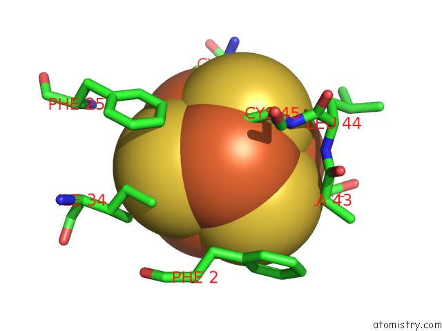
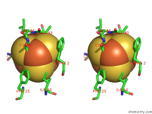
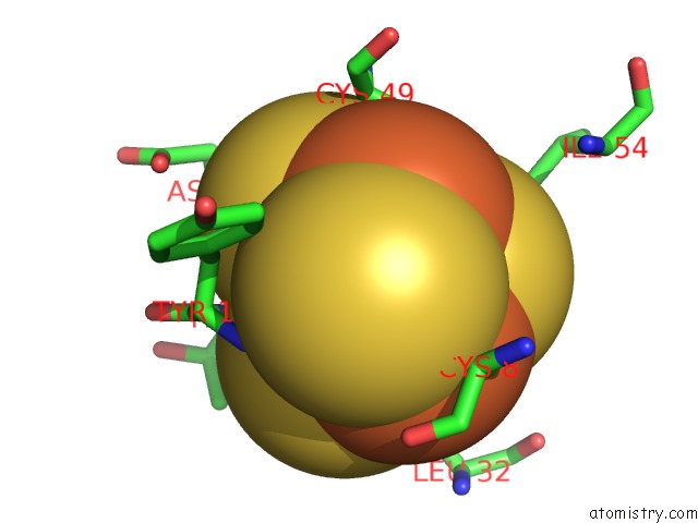
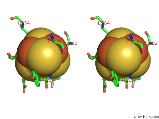
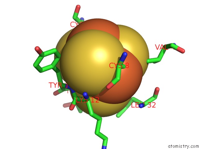
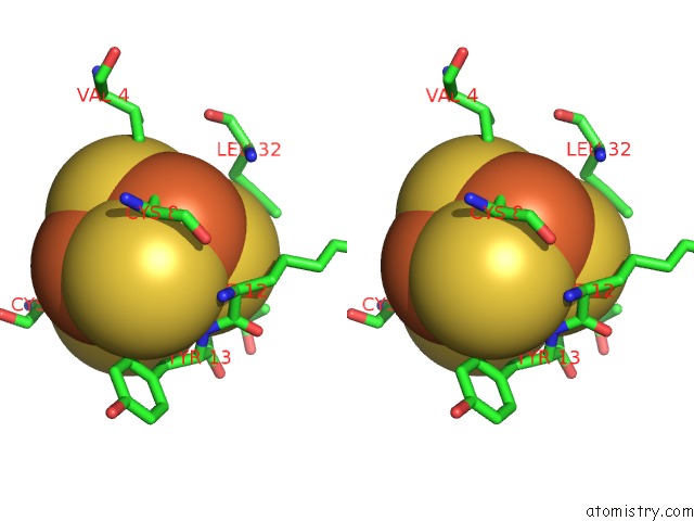
Iron binding site 1 out of 7 in 1pc5
Go back to
Iron binding site 1 out
of 7 in the Crystal Structure of the P50G Mutant of Ferredoxin I at 1.8 A Resolution
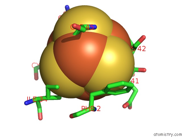
Mono view
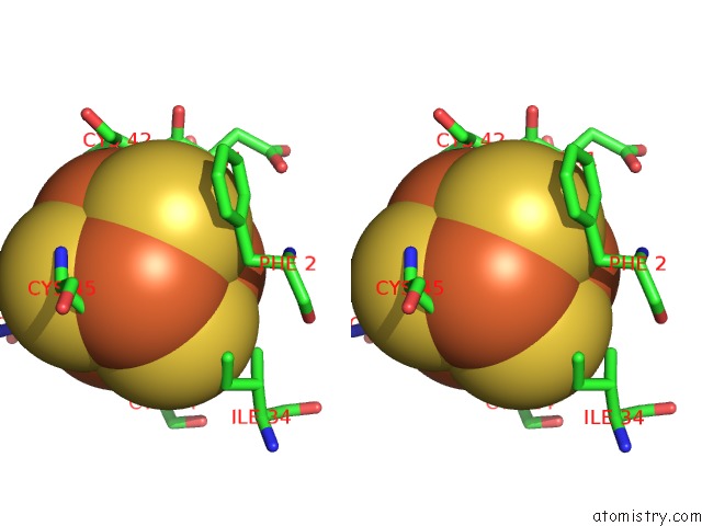
Stereo pair view
Mono view
Stereo pair view
A full contact list of Iron with other atoms in the Fe binding
site number 1 of Crystal Structure of the P50G Mutant of Ferredoxin I at 1.8 A Resolution within 5.0Å range:
|
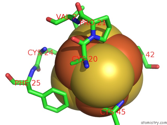
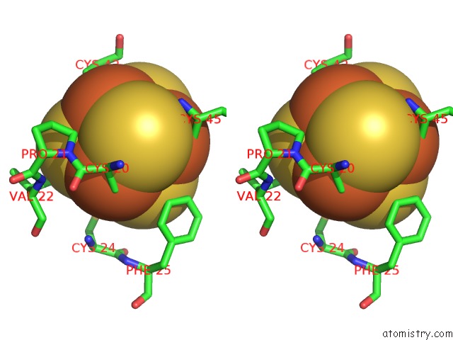
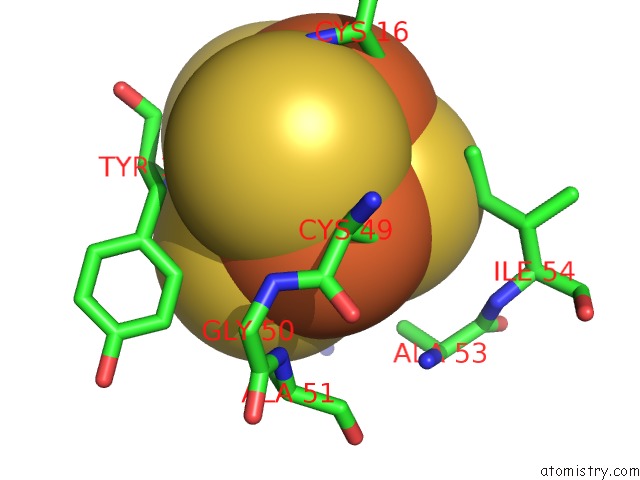
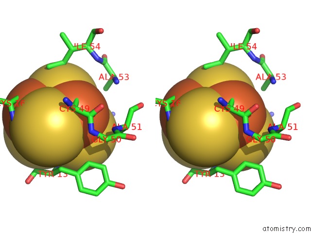
Iron binding site 2 out of 7 in 1pc5
Go back to
Iron binding site 2 out
of 7 in the Crystal Structure of the P50G Mutant of Ferredoxin I at 1.8 A Resolution
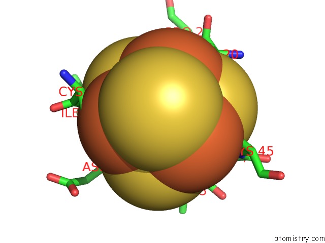
Mono view
Stereo pair view
Mono view
Stereo pair view
A full contact list of Iron with other atoms in the Fe binding
site number 2 of Crystal Structure of the P50G Mutant of Ferredoxin I at 1.8 A Resolution within 5.0Å range:
|
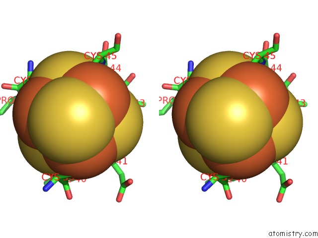
Iron binding site 3 out of 7 in 1pc5
Go back to
Iron binding site 3 out
of 7 in the Crystal Structure of the P50G Mutant of Ferredoxin I at 1.8 A Resolution
Mono view
Stereo pair view
Mono view
Stereo pair view
A full contact list of Iron with other atoms in the Fe binding
site number 3 of Crystal Structure of the P50G Mutant of Ferredoxin I at 1.8 A Resolution within 5.0Å range:
|
Iron binding site 4 out of 7 in 1pc5
Go back to
Iron binding site 4 out
of 7 in the Crystal Structure of the P50G Mutant of Ferredoxin I at 1.8 A Resolution
Mono view
Stereo pair view
Mono view
Stereo pair view
A full contact list of Iron with other atoms in the Fe binding
site number 4 of Crystal Structure of the P50G Mutant of Ferredoxin I at 1.8 A Resolution within 5.0Å range:
|
Iron binding site 5 out of 7 in 1pc5
Go back to
Iron binding site 5 out
of 7 in the Crystal Structure of the P50G Mutant of Ferredoxin I at 1.8 A Resolution
Mono view
Stereo pair view
Mono view
Stereo pair view
A full contact list of Iron with other atoms in the Fe binding
site number 5 of Crystal Structure of the P50G Mutant of Ferredoxin I at 1.8 A Resolution within 5.0Å range:
|
Iron binding site 6 out of 7 in 1pc5
Go back to
Iron binding site 6 out
of 7 in the Crystal Structure of the P50G Mutant of Ferredoxin I at 1.8 A Resolution
Mono view
Stereo pair view
Mono view
Stereo pair view
A full contact list of Iron with other atoms in the Fe binding
site number 6 of Crystal Structure of the P50G Mutant of Ferredoxin I at 1.8 A Resolution within 5.0Å range:
|
Iron binding site 7 out of 7 in 1pc5
Go back to
Iron binding site 7 out
of 7 in the Crystal Structure of the P50G Mutant of Ferredoxin I at 1.8 A Resolution
Mono view
Stereo pair view
Mono view
Stereo pair view
A full contact list of Iron with other atoms in the Fe binding
site number 7 of Crystal Structure of the P50G Mutant of Ferredoxin I at 1.8 A Resolution within 5.0Å range:
|
Reference:
R.Camba,
Y.S.Jung,
L.M.Hunsicker-Wang,
B.K.Burgess,
C.D.Stout,
J.Hirst,
F.A.Armstrong.
Mechanisms of Redox-Coupled Proton Transfer in Proteins: Role of the Proximal Proline in Reactions of the [3FE-4S] Cluster in Azotobacter Vinelandii Ferredoxin I Biochemistry V. 42 10589 2003.
ISSN: ISSN 0006-2960
PubMed: 12962482
DOI: 10.1021/BI035021V
Page generated: Sat Aug 3 13:02:10 2024
ISSN: ISSN 0006-2960
PubMed: 12962482
DOI: 10.1021/BI035021V
Last articles
Zn in 9J0NZn in 9J0O
Zn in 9J0P
Zn in 9FJX
Zn in 9EKB
Zn in 9C0F
Zn in 9CAH
Zn in 9CH0
Zn in 9CH3
Zn in 9CH1