Iron »
PDB 1phb-1q5d »
1phc »
Iron in PDB 1phc: Crystal Structure of Substrate-Free Pseudomonas Putida Cytochrome P450
Enzymatic activity of Crystal Structure of Substrate-Free Pseudomonas Putida Cytochrome P450
All present enzymatic activity of Crystal Structure of Substrate-Free Pseudomonas Putida Cytochrome P450:
1.14.15.1;
1.14.15.1;
Protein crystallography data
The structure of Crystal Structure of Substrate-Free Pseudomonas Putida Cytochrome P450, PDB code: 1phc
was solved by
T.L.Poulos,
with X-Ray Crystallography technique. A brief refinement statistics is given in the table below:
| Resolution Low / High (Å) | N/A / 1.60 |
| Space group | P 21 21 21 |
| Cell size a, b, c (Å), α, β, γ (°) | 108.670, 103.900, 36.380, 90.00, 90.00, 90.00 |
| R / Rfree (%) | n/a / n/a |
Iron Binding Sites:
The binding sites of Iron atom in the Crystal Structure of Substrate-Free Pseudomonas Putida Cytochrome P450
(pdb code 1phc). This binding sites where shown within
5.0 Angstroms radius around Iron atom.
In total only one binding site of Iron was determined in the Crystal Structure of Substrate-Free Pseudomonas Putida Cytochrome P450, PDB code: 1phc:
In total only one binding site of Iron was determined in the Crystal Structure of Substrate-Free Pseudomonas Putida Cytochrome P450, PDB code: 1phc:
Iron binding site 1 out of 1 in 1phc
Go back to
Iron binding site 1 out
of 1 in the Crystal Structure of Substrate-Free Pseudomonas Putida Cytochrome P450
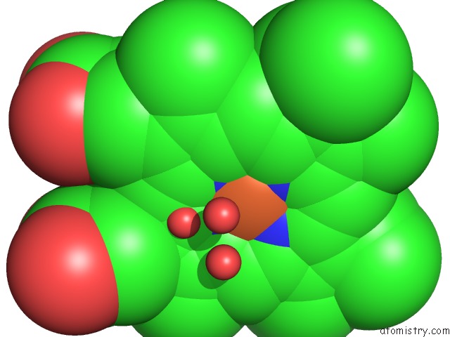
Mono view
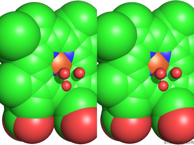
Stereo pair view
Mono view
Stereo pair view
A full contact list of Iron with other atoms in the Fe binding
site number 1 of Crystal Structure of Substrate-Free Pseudomonas Putida Cytochrome P450 within 5.0Å range:
|
Reference:
T.L.Poulos,
B.C.Finzel,
A.J.Howard.
Crystal Structure of Substrate-Free Pseudomonas Putida Cytochrome P-450. Biochemistry V. 25 5314 1986.
ISSN: ISSN 0006-2960
PubMed: 3768350
DOI: 10.1021/BI00366A049
Page generated: Sat Aug 3 13:07:49 2024
ISSN: ISSN 0006-2960
PubMed: 3768350
DOI: 10.1021/BI00366A049
Last articles
Zn in 9JYWZn in 9IR4
Zn in 9IR3
Zn in 9GMX
Zn in 9GMW
Zn in 9JEJ
Zn in 9ERF
Zn in 9ERE
Zn in 9EGV
Zn in 9EGW