Iron »
PDB 1phb-1q5d »
1piz »
Iron in PDB 1piz: Ribonucleotide Reductase R2 D84E Mutant Soaked with Ferrous Ions at Neutral pH
Enzymatic activity of Ribonucleotide Reductase R2 D84E Mutant Soaked with Ferrous Ions at Neutral pH
All present enzymatic activity of Ribonucleotide Reductase R2 D84E Mutant Soaked with Ferrous Ions at Neutral pH:
1.17.4.1;
1.17.4.1;
Protein crystallography data
The structure of Ribonucleotide Reductase R2 D84E Mutant Soaked with Ferrous Ions at Neutral pH, PDB code: 1piz
was solved by
W.C.Voegtli,
M.Sommerhalter,
L.Saleh,
J.Baldwin,
J.M.Bollinger Jr.,
A.C.Rosenzweig,
with X-Ray Crystallography technique. A brief refinement statistics is given in the table below:
| Resolution Low / High (Å) | 23.85 / 1.90 |
| Space group | P 21 21 21 |
| Cell size a, b, c (Å), α, β, γ (°) | 74.000, 84.200, 114.200, 90.00, 90.00, 90.00 |
| R / Rfree (%) | 20.7 / 24.2 |
Other elements in 1piz:
The structure of Ribonucleotide Reductase R2 D84E Mutant Soaked with Ferrous Ions at Neutral pH also contains other interesting chemical elements:
| Mercury | (Hg) | 11 atoms |
Iron Binding Sites:
The binding sites of Iron atom in the Ribonucleotide Reductase R2 D84E Mutant Soaked with Ferrous Ions at Neutral pH
(pdb code 1piz). This binding sites where shown within
5.0 Angstroms radius around Iron atom.
In total 4 binding sites of Iron where determined in the Ribonucleotide Reductase R2 D84E Mutant Soaked with Ferrous Ions at Neutral pH, PDB code: 1piz:
Jump to Iron binding site number: 1; 2; 3; 4;
In total 4 binding sites of Iron where determined in the Ribonucleotide Reductase R2 D84E Mutant Soaked with Ferrous Ions at Neutral pH, PDB code: 1piz:
Jump to Iron binding site number: 1; 2; 3; 4;
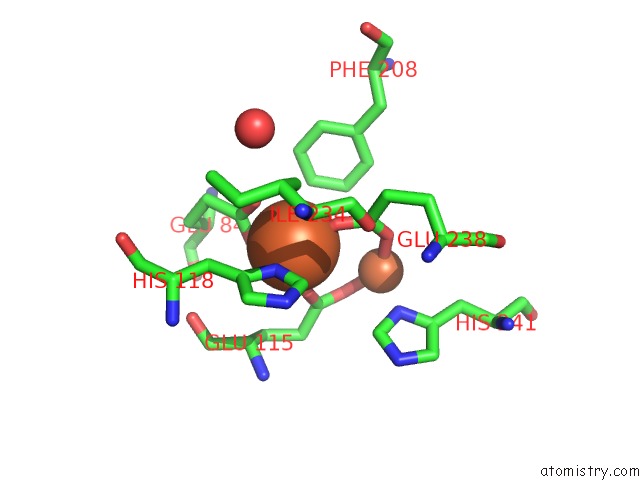
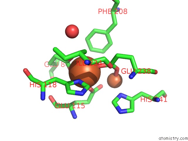
Iron binding site 1 out of 4 in 1piz
Go back to
Iron binding site 1 out
of 4 in the Ribonucleotide Reductase R2 D84E Mutant Soaked with Ferrous Ions at Neutral pH
Mono view
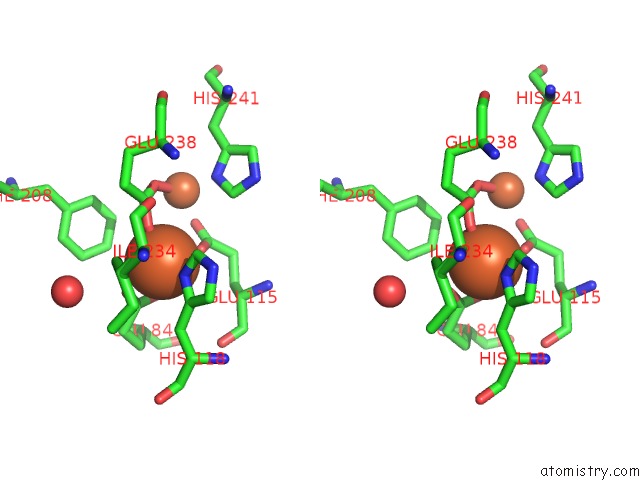
Stereo pair view
Mono view
Stereo pair view
A full contact list of Iron with other atoms in the Fe binding
site number 1 of Ribonucleotide Reductase R2 D84E Mutant Soaked with Ferrous Ions at Neutral pH within 5.0Å range:
|
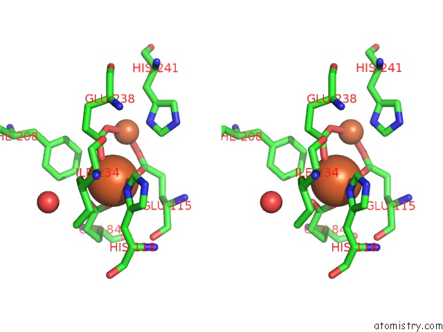
Iron binding site 2 out of 4 in 1piz
Go back to
Iron binding site 2 out
of 4 in the Ribonucleotide Reductase R2 D84E Mutant Soaked with Ferrous Ions at Neutral pH
Mono view
Stereo pair view
Mono view
Stereo pair view
A full contact list of Iron with other atoms in the Fe binding
site number 2 of Ribonucleotide Reductase R2 D84E Mutant Soaked with Ferrous Ions at Neutral pH within 5.0Å range:
|
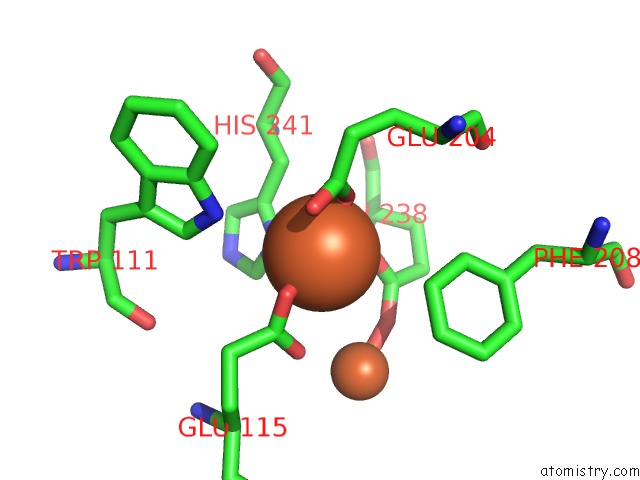
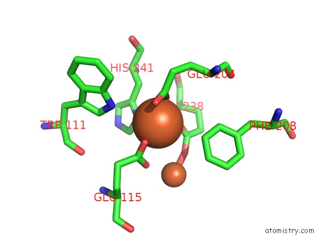
Iron binding site 3 out of 4 in 1piz
Go back to
Iron binding site 3 out
of 4 in the Ribonucleotide Reductase R2 D84E Mutant Soaked with Ferrous Ions at Neutral pH
Mono view
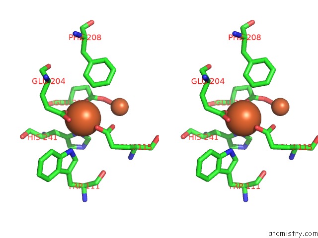
Stereo pair view
Mono view
Stereo pair view
A full contact list of Iron with other atoms in the Fe binding
site number 3 of Ribonucleotide Reductase R2 D84E Mutant Soaked with Ferrous Ions at Neutral pH within 5.0Å range:
|
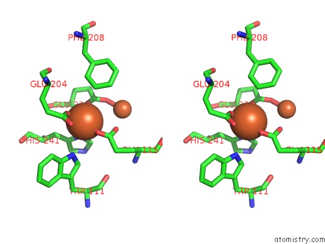
Iron binding site 4 out of 4 in 1piz
Go back to
Iron binding site 4 out
of 4 in the Ribonucleotide Reductase R2 D84E Mutant Soaked with Ferrous Ions at Neutral pH
Mono view
Stereo pair view
Mono view
Stereo pair view
A full contact list of Iron with other atoms in the Fe binding
site number 4 of Ribonucleotide Reductase R2 D84E Mutant Soaked with Ferrous Ions at Neutral pH within 5.0Å range:
|
Reference:
W.C.Voegtli,
M.Sommerhalter,
L.Saleh,
J.Baldwin,
J.M.Bollinger Jr.,
A.C.Rosenzweig.
Variable Coordination Geometries at the Diiron(II) Active Site of Ribonucleotide Reductase R2. J.Am.Chem.Soc. V. 125 15822 2003.
ISSN: ISSN 0002-7863
PubMed: 14677973
DOI: 10.1021/JA0370387
Page generated: Wed Jul 16 19:38:11 2025
ISSN: ISSN 0002-7863
PubMed: 14677973
DOI: 10.1021/JA0370387
Last articles
Fe in 2YXOFe in 2YRS
Fe in 2YXC
Fe in 2YNM
Fe in 2YVJ
Fe in 2YP1
Fe in 2YU2
Fe in 2YU1
Fe in 2YQB
Fe in 2YOO