Iron »
PDB 1phb-1q5d »
1pj0 »
Iron in PDB 1pj0: Ribonucleotide Reductase R2-D84E/W48F Mutant Soaked with Ferrous Ions at Neutral pH
Enzymatic activity of Ribonucleotide Reductase R2-D84E/W48F Mutant Soaked with Ferrous Ions at Neutral pH
All present enzymatic activity of Ribonucleotide Reductase R2-D84E/W48F Mutant Soaked with Ferrous Ions at Neutral pH:
1.17.4.1;
1.17.4.1;
Protein crystallography data
The structure of Ribonucleotide Reductase R2-D84E/W48F Mutant Soaked with Ferrous Ions at Neutral pH, PDB code: 1pj0
was solved by
W.C.Voegtli,
M.Sommerhalter,
L.Saleh,
J.Baldwin,
J.M.Bollinger Jr.,
A.C.Rosenzweig,
with X-Ray Crystallography technique. A brief refinement statistics is given in the table below:
| Resolution Low / High (Å) | 19.32 / 1.90 |
| Space group | P 21 21 21 |
| Cell size a, b, c (Å), α, β, γ (°) | 74.000, 84.400, 114.600, 90.00, 90.00, 90.00 |
| R / Rfree (%) | 20.4 / 23.6 |
Other elements in 1pj0:
The structure of Ribonucleotide Reductase R2-D84E/W48F Mutant Soaked with Ferrous Ions at Neutral pH also contains other interesting chemical elements:
| Mercury | (Hg) | 9 atoms |
Iron Binding Sites:
The binding sites of Iron atom in the Ribonucleotide Reductase R2-D84E/W48F Mutant Soaked with Ferrous Ions at Neutral pH
(pdb code 1pj0). This binding sites where shown within
5.0 Angstroms radius around Iron atom.
In total 4 binding sites of Iron where determined in the Ribonucleotide Reductase R2-D84E/W48F Mutant Soaked with Ferrous Ions at Neutral pH, PDB code: 1pj0:
Jump to Iron binding site number: 1; 2; 3; 4;
In total 4 binding sites of Iron where determined in the Ribonucleotide Reductase R2-D84E/W48F Mutant Soaked with Ferrous Ions at Neutral pH, PDB code: 1pj0:
Jump to Iron binding site number: 1; 2; 3; 4;
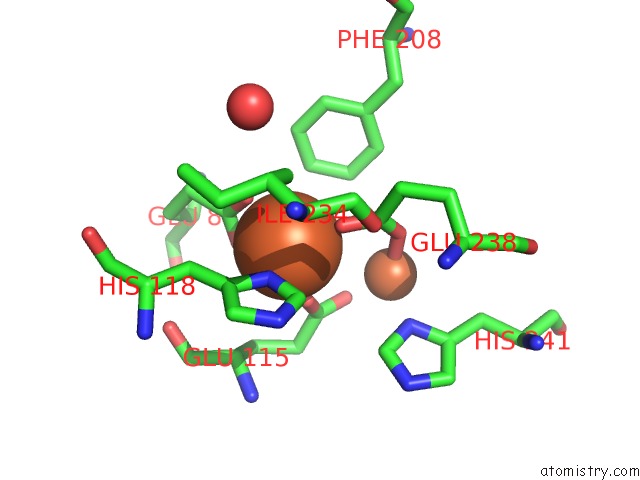
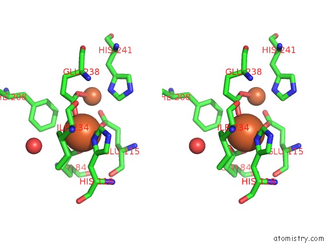
Iron binding site 1 out of 4 in 1pj0
Go back to
Iron binding site 1 out
of 4 in the Ribonucleotide Reductase R2-D84E/W48F Mutant Soaked with Ferrous Ions at Neutral pH
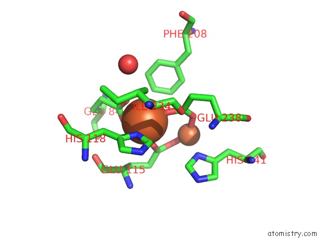
Mono view
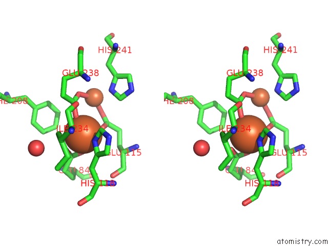
Stereo pair view
Mono view
Stereo pair view
A full contact list of Iron with other atoms in the Fe binding
site number 1 of Ribonucleotide Reductase R2-D84E/W48F Mutant Soaked with Ferrous Ions at Neutral pH within 5.0Å range:
|
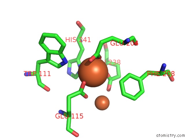
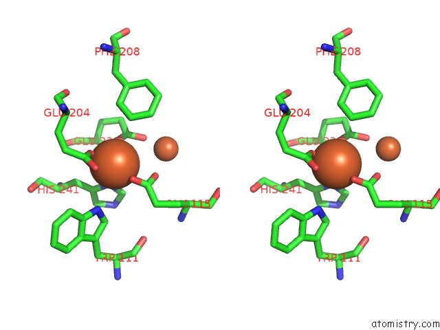
Iron binding site 2 out of 4 in 1pj0
Go back to
Iron binding site 2 out
of 4 in the Ribonucleotide Reductase R2-D84E/W48F Mutant Soaked with Ferrous Ions at Neutral pH
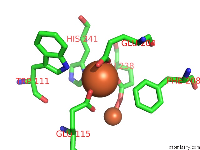
Mono view
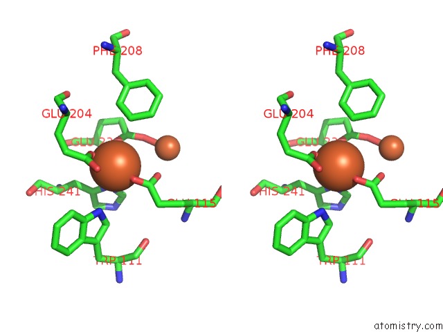
Stereo pair view
Mono view
Stereo pair view
A full contact list of Iron with other atoms in the Fe binding
site number 2 of Ribonucleotide Reductase R2-D84E/W48F Mutant Soaked with Ferrous Ions at Neutral pH within 5.0Å range:
|
Iron binding site 3 out of 4 in 1pj0
Go back to
Iron binding site 3 out
of 4 in the Ribonucleotide Reductase R2-D84E/W48F Mutant Soaked with Ferrous Ions at Neutral pH
Mono view
Stereo pair view
Mono view
Stereo pair view
A full contact list of Iron with other atoms in the Fe binding
site number 3 of Ribonucleotide Reductase R2-D84E/W48F Mutant Soaked with Ferrous Ions at Neutral pH within 5.0Å range:
|
Iron binding site 4 out of 4 in 1pj0
Go back to
Iron binding site 4 out
of 4 in the Ribonucleotide Reductase R2-D84E/W48F Mutant Soaked with Ferrous Ions at Neutral pH
Mono view
Stereo pair view
Mono view
Stereo pair view
A full contact list of Iron with other atoms in the Fe binding
site number 4 of Ribonucleotide Reductase R2-D84E/W48F Mutant Soaked with Ferrous Ions at Neutral pH within 5.0Å range:
|
Reference:
W.C.Voegtli,
M.Sommerhalter,
L.Saleh,
J.Baldwin,
J.M.Bollinger Jr.,
A.C.Rosenzweig.
Variable Coordination Geometries at the Diiron(II) Active Site of Ribonucleotide Reductase R2. J.Am.Chem.Soc. V. 125 15822 2003.
ISSN: ISSN 0002-7863
PubMed: 14677973
DOI: 10.1021/JA0370387
Page generated: Sat Aug 3 13:08:59 2024
ISSN: ISSN 0002-7863
PubMed: 14677973
DOI: 10.1021/JA0370387
Last articles
F in 4PX5F in 4PY4
F in 4PWL
F in 4PVU
F in 4PUW
F in 4PU9
F in 4PUU
F in 4PPP
F in 4PTW
F in 4PP7