Iron »
PDB 1phb-1q5d »
1pmb »
Iron in PDB 1pmb: The Determination of the Crystal Structure of Recombinant Pig Myoglobin By Molecular Replacement and Its Refinement
Protein crystallography data
The structure of The Determination of the Crystal Structure of Recombinant Pig Myoglobin By Molecular Replacement and Its Refinement, PDB code: 1pmb
was solved by
S.J.Smerdon,
T.J.Oldfield,
E.J.Dodson,
G.G.Dodson,
R.E.Hubbard,
A.J.Wilkinson,
with X-Ray Crystallography technique. A brief refinement statistics is given in the table below:
| Resolution Low / High (Å) | 10.00 / 2.50 |
| Space group | I 21 |
| Cell size a, b, c (Å), α, β, γ (°) | 124.910, 42.630, 92.200, 90.00, 92.16, 90.00 |
| R / Rfree (%) | n/a / n/a |
Iron Binding Sites:
The binding sites of Iron atom in the The Determination of the Crystal Structure of Recombinant Pig Myoglobin By Molecular Replacement and Its Refinement
(pdb code 1pmb). This binding sites where shown within
5.0 Angstroms radius around Iron atom.
In total 2 binding sites of Iron where determined in the The Determination of the Crystal Structure of Recombinant Pig Myoglobin By Molecular Replacement and Its Refinement, PDB code: 1pmb:
Jump to Iron binding site number: 1; 2;
In total 2 binding sites of Iron where determined in the The Determination of the Crystal Structure of Recombinant Pig Myoglobin By Molecular Replacement and Its Refinement, PDB code: 1pmb:
Jump to Iron binding site number: 1; 2;
Iron binding site 1 out of 2 in 1pmb
Go back to
Iron binding site 1 out
of 2 in the The Determination of the Crystal Structure of Recombinant Pig Myoglobin By Molecular Replacement and Its Refinement
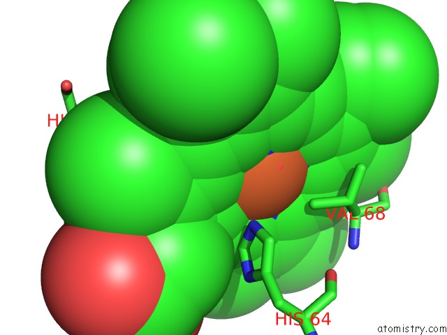
Mono view
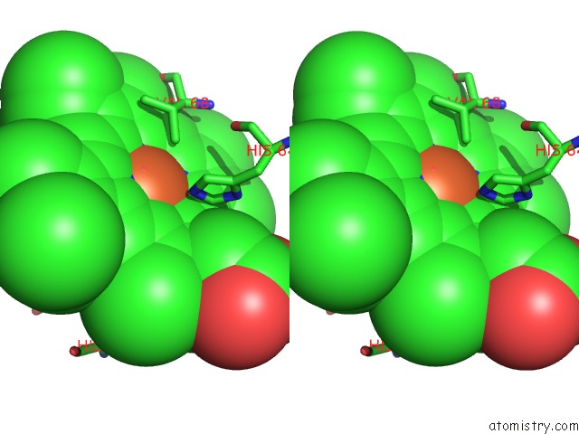
Stereo pair view
Mono view
Stereo pair view
A full contact list of Iron with other atoms in the Fe binding
site number 1 of The Determination of the Crystal Structure of Recombinant Pig Myoglobin By Molecular Replacement and Its Refinement within 5.0Å range:
|
Iron binding site 2 out of 2 in 1pmb
Go back to
Iron binding site 2 out
of 2 in the The Determination of the Crystal Structure of Recombinant Pig Myoglobin By Molecular Replacement and Its Refinement
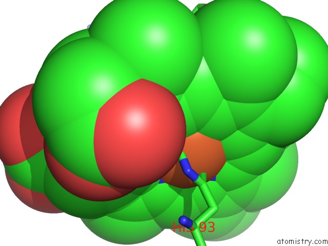
Mono view
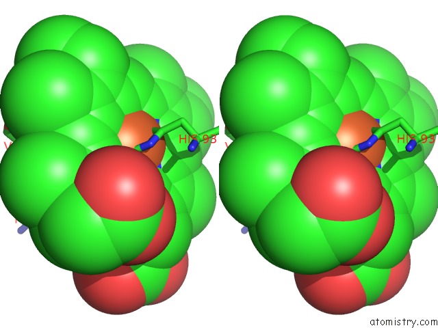
Stereo pair view
Mono view
Stereo pair view
A full contact list of Iron with other atoms in the Fe binding
site number 2 of The Determination of the Crystal Structure of Recombinant Pig Myoglobin By Molecular Replacement and Its Refinement within 5.0Å range:
|
Reference:
S.J.Smerdon,
T.J.Oldfield,
E.J.Dodson,
G.G.Dodson,
R.E.Hubbard,
A.J.Wilkinson.
Determination of the Crystal Structure of Recombinant Pig Myoglobin By Molecular Replacement and Its Refinement. Acta Crystallogr.,Sect.B V. 46 370 1990.
ISSN: ISSN 0108-7681
PubMed: 2383370
DOI: 10.1107/S0108768189012450
Page generated: Sat Aug 3 13:11:21 2024
ISSN: ISSN 0108-7681
PubMed: 2383370
DOI: 10.1107/S0108768189012450
Last articles
Zn in 9J0NZn in 9J0O
Zn in 9J0P
Zn in 9FJX
Zn in 9EKB
Zn in 9C0F
Zn in 9CAH
Zn in 9CH0
Zn in 9CH3
Zn in 9CH1