Iron »
PDB 1phb-1q5d »
1pth »
Iron in PDB 1pth: The Structural Basis of Aspirin Activity Inferred From the Crystal Structure of Inactivated Prostaglandin H2 Synthase
Enzymatic activity of The Structural Basis of Aspirin Activity Inferred From the Crystal Structure of Inactivated Prostaglandin H2 Synthase
All present enzymatic activity of The Structural Basis of Aspirin Activity Inferred From the Crystal Structure of Inactivated Prostaglandin H2 Synthase:
1.14.99.1;
1.14.99.1;
Protein crystallography data
The structure of The Structural Basis of Aspirin Activity Inferred From the Crystal Structure of Inactivated Prostaglandin H2 Synthase, PDB code: 1pth
was solved by
P.J.Loll,
D.Picot,
R.M.Garavito,
with X-Ray Crystallography technique. A brief refinement statistics is given in the table below:
| Resolution Low / High (Å) | 8.00 / 3.40 |
| Space group | I 2 2 2 |
| Cell size a, b, c (Å), α, β, γ (°) | 99.570, 209.800, 235.000, 90.00, 90.00, 90.00 |
| R / Rfree (%) | 18.6 / 22.7 |
Other elements in 1pth:
The structure of The Structural Basis of Aspirin Activity Inferred From the Crystal Structure of Inactivated Prostaglandin H2 Synthase also contains other interesting chemical elements:
| Bromine | (Br) | 2 atoms |
Iron Binding Sites:
The binding sites of Iron atom in the The Structural Basis of Aspirin Activity Inferred From the Crystal Structure of Inactivated Prostaglandin H2 Synthase
(pdb code 1pth). This binding sites where shown within
5.0 Angstroms radius around Iron atom.
In total 2 binding sites of Iron where determined in the The Structural Basis of Aspirin Activity Inferred From the Crystal Structure of Inactivated Prostaglandin H2 Synthase, PDB code: 1pth:
Jump to Iron binding site number: 1; 2;
In total 2 binding sites of Iron where determined in the The Structural Basis of Aspirin Activity Inferred From the Crystal Structure of Inactivated Prostaglandin H2 Synthase, PDB code: 1pth:
Jump to Iron binding site number: 1; 2;
Iron binding site 1 out of 2 in 1pth
Go back to
Iron binding site 1 out
of 2 in the The Structural Basis of Aspirin Activity Inferred From the Crystal Structure of Inactivated Prostaglandin H2 Synthase
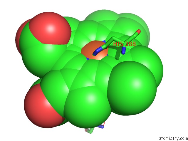
Mono view
Stereo pair view
Mono view
Stereo pair view
A full contact list of Iron with other atoms in the Fe binding
site number 1 of The Structural Basis of Aspirin Activity Inferred From the Crystal Structure of Inactivated Prostaglandin H2 Synthase within 5.0Å range:
|
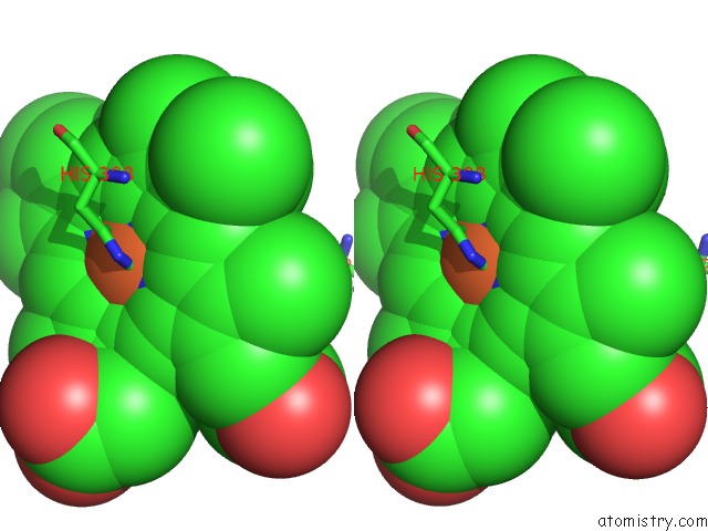
Iron binding site 2 out of 2 in 1pth
Go back to
Iron binding site 2 out
of 2 in the The Structural Basis of Aspirin Activity Inferred From the Crystal Structure of Inactivated Prostaglandin H2 Synthase

Mono view

Stereo pair view
Mono view
Stereo pair view
A full contact list of Iron with other atoms in the Fe binding
site number 2 of The Structural Basis of Aspirin Activity Inferred From the Crystal Structure of Inactivated Prostaglandin H2 Synthase within 5.0Å range:
|
Reference:
P.J.Loll,
D.Picot,
R.M.Garavito.
The Structural Basis of Aspirin Activity Inferred From the Crystal Structure of Inactivated Prostaglandin H2 Synthase. Nat.Struct.Biol. V. 2 637 1995.
ISSN: ISSN 1072-8368
PubMed: 7552725
DOI: 10.1038/NSB0895-637
Page generated: Sat Aug 3 13:13:44 2024
ISSN: ISSN 1072-8368
PubMed: 7552725
DOI: 10.1038/NSB0895-637
Last articles
Zn in 9JYWZn in 9IR4
Zn in 9IR3
Zn in 9GMX
Zn in 9GMW
Zn in 9JEJ
Zn in 9ERF
Zn in 9ERE
Zn in 9EGV
Zn in 9EGW