Iron »
PDB 1phb-1q5d »
1pxx »
Iron in PDB 1pxx: Crystal Structure of Diclofenac Bound to the Cyclooxygenase Active Site of Cox-2
Enzymatic activity of Crystal Structure of Diclofenac Bound to the Cyclooxygenase Active Site of Cox-2
All present enzymatic activity of Crystal Structure of Diclofenac Bound to the Cyclooxygenase Active Site of Cox-2:
1.14.99.1;
1.14.99.1;
Protein crystallography data
The structure of Crystal Structure of Diclofenac Bound to the Cyclooxygenase Active Site of Cox-2, PDB code: 1pxx
was solved by
J.R.Kiefer,
S.W.Rowlinson,
J.J.Prusakiewicz,
J.L.Pawlitz,
K.R.Kozak,
A.S.Kalgutkar,
W.C.Stallings,
L.J.Marnett,
R.G.Kurumbail,
with X-Ray Crystallography technique. A brief refinement statistics is given in the table below:
| Resolution Low / High (Å) | 20.00 / 2.90 |
| Space group | P 21 21 2 |
| Cell size a, b, c (Å), α, β, γ (°) | 181.145, 135.090, 124.165, 90.00, 90.00, 90.00 |
| R / Rfree (%) | 25.4 / 30.2 |
Other elements in 1pxx:
The structure of Crystal Structure of Diclofenac Bound to the Cyclooxygenase Active Site of Cox-2 also contains other interesting chemical elements:
| Chlorine | (Cl) | 8 atoms |
Iron Binding Sites:
The binding sites of Iron atom in the Crystal Structure of Diclofenac Bound to the Cyclooxygenase Active Site of Cox-2
(pdb code 1pxx). This binding sites where shown within
5.0 Angstroms radius around Iron atom.
In total 4 binding sites of Iron where determined in the Crystal Structure of Diclofenac Bound to the Cyclooxygenase Active Site of Cox-2, PDB code: 1pxx:
Jump to Iron binding site number: 1; 2; 3; 4;
In total 4 binding sites of Iron where determined in the Crystal Structure of Diclofenac Bound to the Cyclooxygenase Active Site of Cox-2, PDB code: 1pxx:
Jump to Iron binding site number: 1; 2; 3; 4;
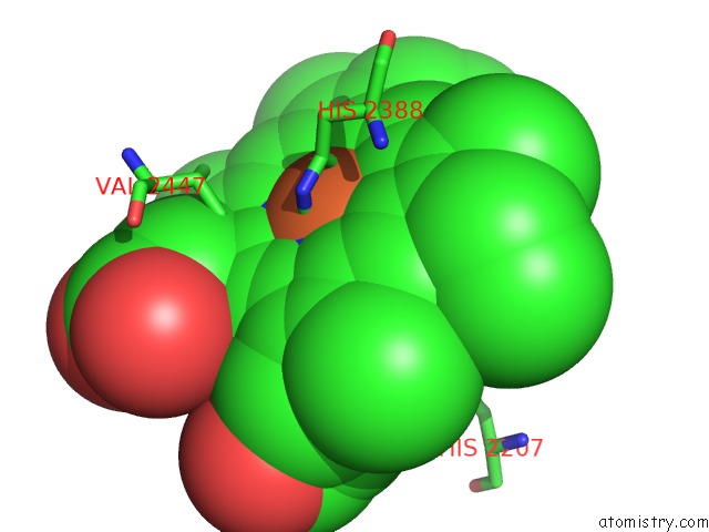
Iron binding site 1 out of 4 in 1pxx
Go back to
Iron binding site 1 out
of 4 in the Crystal Structure of Diclofenac Bound to the Cyclooxygenase Active Site of Cox-2
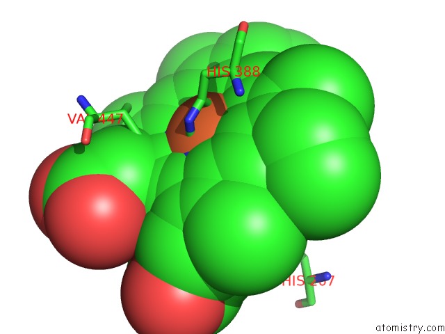
Mono view
Stereo pair view
Mono view
Stereo pair view
A full contact list of Iron with other atoms in the Fe binding
site number 1 of Crystal Structure of Diclofenac Bound to the Cyclooxygenase Active Site of Cox-2 within 5.0Å range:
|
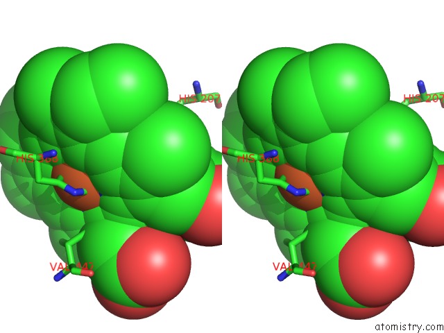
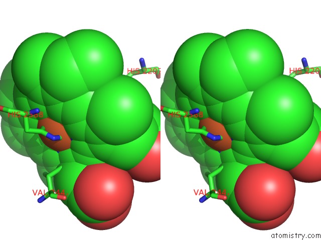
Iron binding site 2 out of 4 in 1pxx
Go back to
Iron binding site 2 out
of 4 in the Crystal Structure of Diclofenac Bound to the Cyclooxygenase Active Site of Cox-2
Mono view
Stereo pair view
Mono view
Stereo pair view
A full contact list of Iron with other atoms in the Fe binding
site number 2 of Crystal Structure of Diclofenac Bound to the Cyclooxygenase Active Site of Cox-2 within 5.0Å range:
|
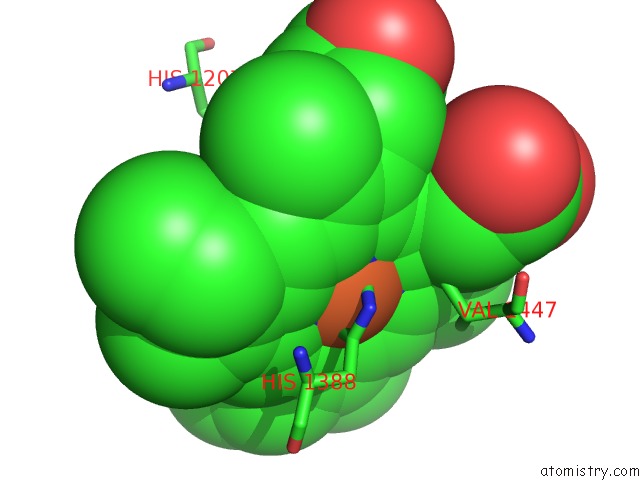
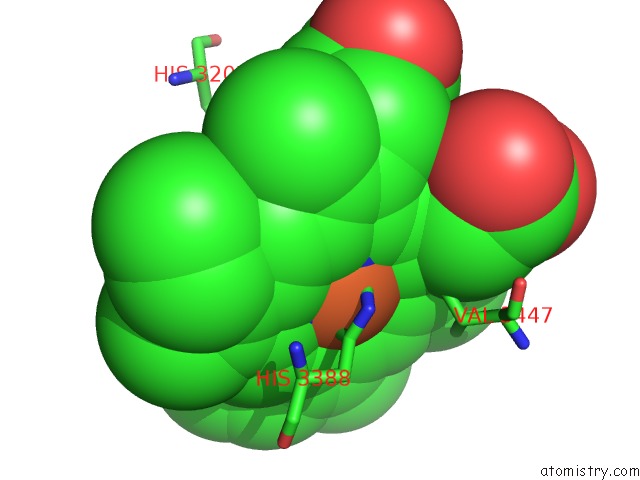
Iron binding site 3 out of 4 in 1pxx
Go back to
Iron binding site 3 out
of 4 in the Crystal Structure of Diclofenac Bound to the Cyclooxygenase Active Site of Cox-2
Mono view
Stereo pair view
Mono view
Stereo pair view
A full contact list of Iron with other atoms in the Fe binding
site number 3 of Crystal Structure of Diclofenac Bound to the Cyclooxygenase Active Site of Cox-2 within 5.0Å range:
|
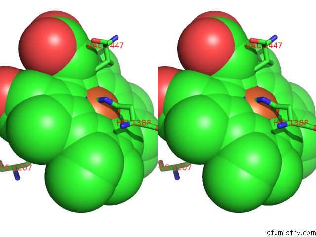
Iron binding site 4 out of 4 in 1pxx
Go back to
Iron binding site 4 out
of 4 in the Crystal Structure of Diclofenac Bound to the Cyclooxygenase Active Site of Cox-2
Mono view
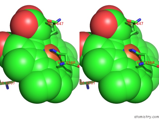
Stereo pair view
Mono view
Stereo pair view
A full contact list of Iron with other atoms in the Fe binding
site number 4 of Crystal Structure of Diclofenac Bound to the Cyclooxygenase Active Site of Cox-2 within 5.0Å range:
|
Reference:
S.W.Rowlinson,
J.R.Kiefer,
J.J.Prusakiewicz,
J.L.Pawlitz,
K.R.Kozak,
A.S.Kalgutkar,
W.C.Stallings,
R.G.Kurumbail,
L.J.Marnett.
A Novel Mechanism of Cyclooxygenase-2 Inhibition Involving Interactions with Ser-530 and Tyr-385. J.Biol.Chem. V. 278 45763 2003.
ISSN: ISSN 0021-9258
PubMed: 12925531
DOI: 10.1074/JBC.M305481200
Page generated: Sat Aug 3 13:14:16 2024
ISSN: ISSN 0021-9258
PubMed: 12925531
DOI: 10.1074/JBC.M305481200
Last articles
Zn in 9J0NZn in 9J0O
Zn in 9J0P
Zn in 9FJX
Zn in 9EKB
Zn in 9C0F
Zn in 9CAH
Zn in 9CH0
Zn in 9CH3
Zn in 9CH1