Iron »
PDB 1phb-1q5d »
1q16 »
Iron in PDB 1q16: Crystal Structure of Nitrate Reductase A, Narghi, From Escherichia Coli
Enzymatic activity of Crystal Structure of Nitrate Reductase A, Narghi, From Escherichia Coli
All present enzymatic activity of Crystal Structure of Nitrate Reductase A, Narghi, From Escherichia Coli:
1.7.99.4;
1.7.99.4;
Protein crystallography data
The structure of Crystal Structure of Nitrate Reductase A, Narghi, From Escherichia Coli, PDB code: 1q16
was solved by
M.G.Bertero,
N.C.J.Strynadka,
with X-Ray Crystallography technique. A brief refinement statistics is given in the table below:
| Resolution Low / High (Å) | 29.68 / 1.90 |
| Space group | C 2 2 21 |
| Cell size a, b, c (Å), α, β, γ (°) | 154.175, 241.376, 139.494, 90.00, 90.00, 90.00 |
| R / Rfree (%) | 20.2 / 23 |
Other elements in 1q16:
The structure of Crystal Structure of Nitrate Reductase A, Narghi, From Escherichia Coli also contains other interesting chemical elements:
| Molybdenum | (Mo) | 1 atom |
Iron Binding Sites:
Pages:
>>> Page 1 <<< Page 2, Binding sites: 11 - 20; Page 3, Binding sites: 21 - 21;Binding sites:
The binding sites of Iron atom in the Crystal Structure of Nitrate Reductase A, Narghi, From Escherichia Coli (pdb code 1q16). This binding sites where shown within 5.0 Angstroms radius around Iron atom.In total 21 binding sites of Iron where determined in the Crystal Structure of Nitrate Reductase A, Narghi, From Escherichia Coli, PDB code: 1q16:
Jump to Iron binding site number: 1; 2; 3; 4; 5; 6; 7; 8; 9; 10;
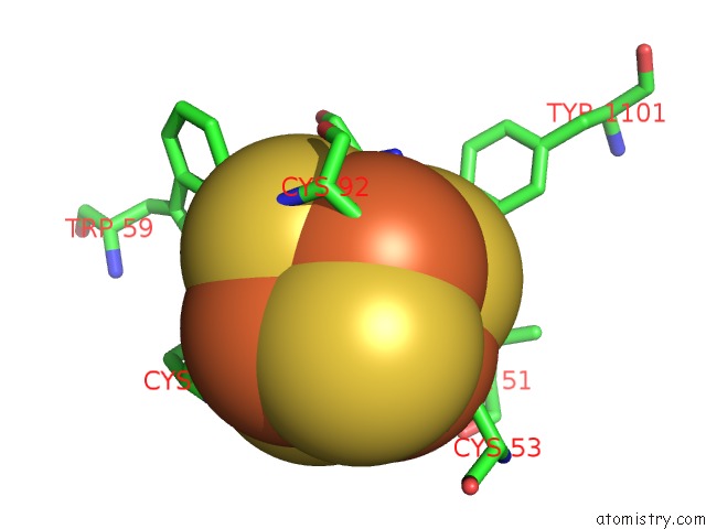
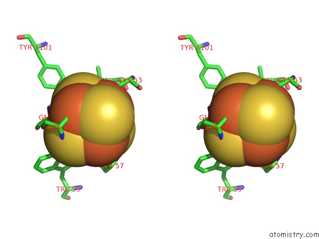
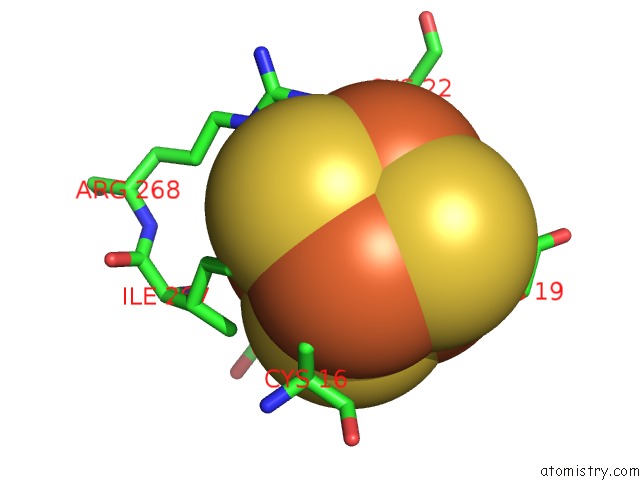
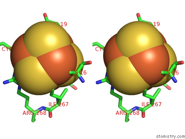
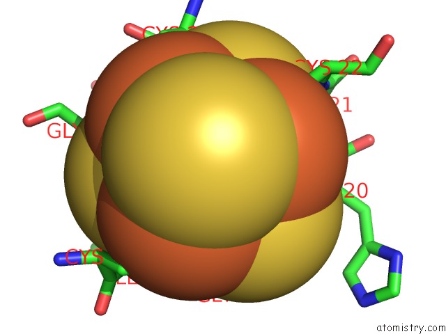
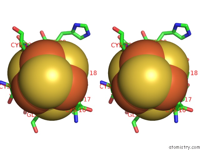
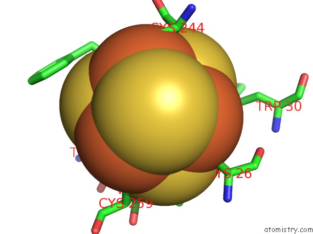
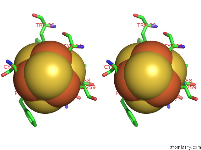
Iron binding site 1 out of 21 in 1q16
Go back to
Iron binding site 1 out
of 21 in the Crystal Structure of Nitrate Reductase A, Narghi, From Escherichia Coli
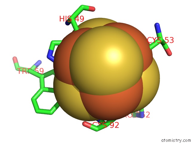
Mono view
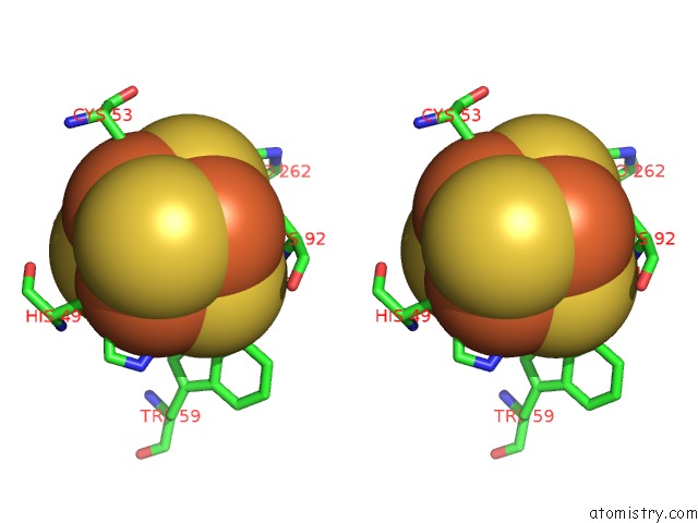
Stereo pair view
Mono view
Stereo pair view
A full contact list of Iron with other atoms in the Fe binding
site number 1 of Crystal Structure of Nitrate Reductase A, Narghi, From Escherichia Coli within 5.0Å range:
|
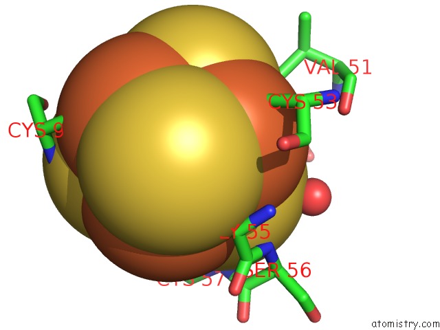
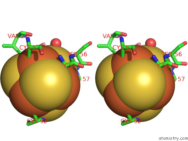
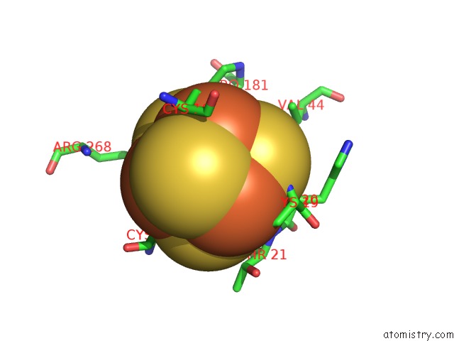
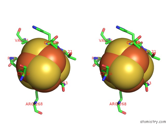
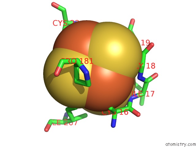
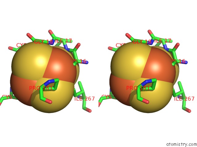
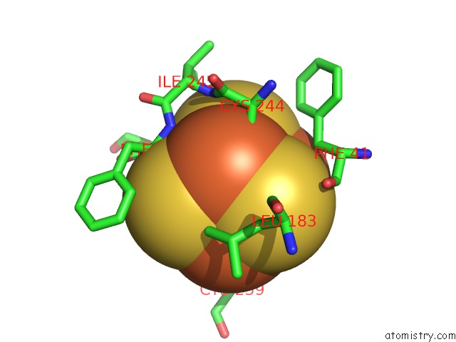
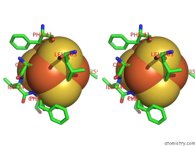
Iron binding site 2 out of 21 in 1q16
Go back to
Iron binding site 2 out
of 21 in the Crystal Structure of Nitrate Reductase A, Narghi, From Escherichia Coli
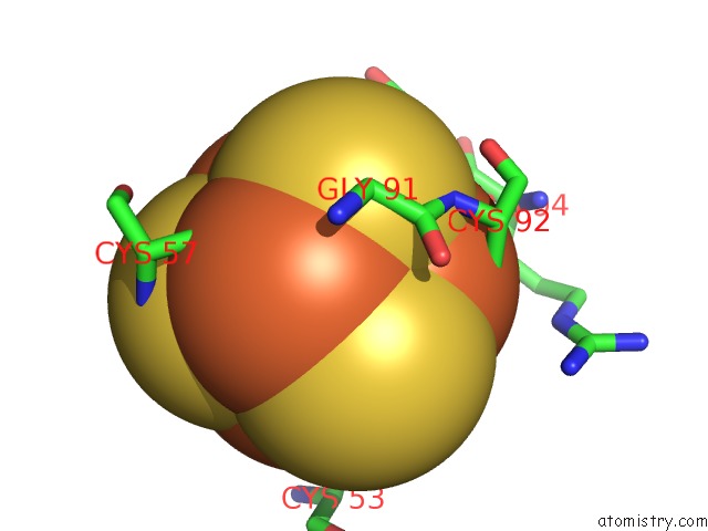
Mono view
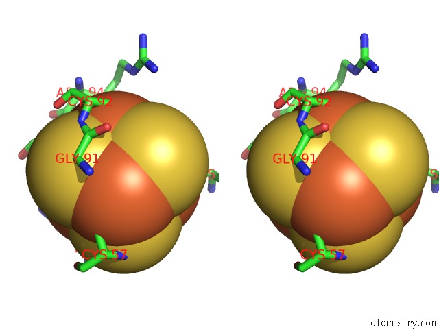
Stereo pair view
Mono view
Stereo pair view
A full contact list of Iron with other atoms in the Fe binding
site number 2 of Crystal Structure of Nitrate Reductase A, Narghi, From Escherichia Coli within 5.0Å range:
|
Iron binding site 3 out of 21 in 1q16
Go back to
Iron binding site 3 out
of 21 in the Crystal Structure of Nitrate Reductase A, Narghi, From Escherichia Coli
Mono view
Stereo pair view
Mono view
Stereo pair view
A full contact list of Iron with other atoms in the Fe binding
site number 3 of Crystal Structure of Nitrate Reductase A, Narghi, From Escherichia Coli within 5.0Å range:
|
Iron binding site 4 out of 21 in 1q16
Go back to
Iron binding site 4 out
of 21 in the Crystal Structure of Nitrate Reductase A, Narghi, From Escherichia Coli
Mono view
Stereo pair view
Mono view
Stereo pair view
A full contact list of Iron with other atoms in the Fe binding
site number 4 of Crystal Structure of Nitrate Reductase A, Narghi, From Escherichia Coli within 5.0Å range:
|
Iron binding site 5 out of 21 in 1q16
Go back to
Iron binding site 5 out
of 21 in the Crystal Structure of Nitrate Reductase A, Narghi, From Escherichia Coli
Mono view
Stereo pair view
Mono view
Stereo pair view
A full contact list of Iron with other atoms in the Fe binding
site number 5 of Crystal Structure of Nitrate Reductase A, Narghi, From Escherichia Coli within 5.0Å range:
|
Iron binding site 6 out of 21 in 1q16
Go back to
Iron binding site 6 out
of 21 in the Crystal Structure of Nitrate Reductase A, Narghi, From Escherichia Coli
Mono view
Stereo pair view
Mono view
Stereo pair view
A full contact list of Iron with other atoms in the Fe binding
site number 6 of Crystal Structure of Nitrate Reductase A, Narghi, From Escherichia Coli within 5.0Å range:
|
Iron binding site 7 out of 21 in 1q16
Go back to
Iron binding site 7 out
of 21 in the Crystal Structure of Nitrate Reductase A, Narghi, From Escherichia Coli
Mono view
Stereo pair view
Mono view
Stereo pair view
A full contact list of Iron with other atoms in the Fe binding
site number 7 of Crystal Structure of Nitrate Reductase A, Narghi, From Escherichia Coli within 5.0Å range:
|
Iron binding site 8 out of 21 in 1q16
Go back to
Iron binding site 8 out
of 21 in the Crystal Structure of Nitrate Reductase A, Narghi, From Escherichia Coli
Mono view
Stereo pair view
Mono view
Stereo pair view
A full contact list of Iron with other atoms in the Fe binding
site number 8 of Crystal Structure of Nitrate Reductase A, Narghi, From Escherichia Coli within 5.0Å range:
|
Iron binding site 9 out of 21 in 1q16
Go back to
Iron binding site 9 out
of 21 in the Crystal Structure of Nitrate Reductase A, Narghi, From Escherichia Coli
Mono view
Stereo pair view
Mono view
Stereo pair view
A full contact list of Iron with other atoms in the Fe binding
site number 9 of Crystal Structure of Nitrate Reductase A, Narghi, From Escherichia Coli within 5.0Å range:
|
Iron binding site 10 out of 21 in 1q16
Go back to
Iron binding site 10 out
of 21 in the Crystal Structure of Nitrate Reductase A, Narghi, From Escherichia Coli
Mono view
Stereo pair view
Mono view
Stereo pair view
A full contact list of Iron with other atoms in the Fe binding
site number 10 of Crystal Structure of Nitrate Reductase A, Narghi, From Escherichia Coli within 5.0Å range:
|
Reference:
M.G.Bertero,
R.A.Rothery,
M.Palak,
C.Hou,
D.Lim,
F.Blasco,
J.H.Weiner,
N.C.J.Strynadka.
Insights Into the Respiratory Electron Transfer Pathway From the Structure of Nitrate Reductase A Nat.Struct.Biol. V. 10 681 2003.
ISSN: ISSN 1072-8368
PubMed: 12910261
DOI: 10.1038/NSB969
Page generated: Sat Aug 3 13:15:00 2024
ISSN: ISSN 1072-8368
PubMed: 12910261
DOI: 10.1038/NSB969
Last articles
Zn in 9MJ5Zn in 9HNW
Zn in 9G0L
Zn in 9FNE
Zn in 9DZN
Zn in 9E0I
Zn in 9D32
Zn in 9DAK
Zn in 8ZXC
Zn in 8ZUF