Iron »
PDB 1phb-1q5d »
1q4g »
Iron in PDB 1q4g: 2.0 Angstrom Crystal Structure of Ovine Prostaglandin H2 Synthase-1, in Complex with Alpha-Methyl-4-Biphenylacetic Acid
Enzymatic activity of 2.0 Angstrom Crystal Structure of Ovine Prostaglandin H2 Synthase-1, in Complex with Alpha-Methyl-4-Biphenylacetic Acid
All present enzymatic activity of 2.0 Angstrom Crystal Structure of Ovine Prostaglandin H2 Synthase-1, in Complex with Alpha-Methyl-4-Biphenylacetic Acid:
1.14.99.1;
1.14.99.1;
Protein crystallography data
The structure of 2.0 Angstrom Crystal Structure of Ovine Prostaglandin H2 Synthase-1, in Complex with Alpha-Methyl-4-Biphenylacetic Acid, PDB code: 1q4g
was solved by
K.Gupta,
B.S.Selinksy,
C.J.Kaub,
A.K.Katz,
P.J.Loll,
with X-Ray Crystallography technique. A brief refinement statistics is given in the table below:
| Resolution Low / High (Å) | 43.68 / 2.00 |
| Space group | I 2 2 2 |
| Cell size a, b, c (Å), α, β, γ (°) | 98.147, 203.859, 223.599, 90.00, 90.00, 90.00 |
| R / Rfree (%) | 21.7 / 23.1 |
Iron Binding Sites:
The binding sites of Iron atom in the 2.0 Angstrom Crystal Structure of Ovine Prostaglandin H2 Synthase-1, in Complex with Alpha-Methyl-4-Biphenylacetic Acid
(pdb code 1q4g). This binding sites where shown within
5.0 Angstroms radius around Iron atom.
In total 4 binding sites of Iron where determined in the 2.0 Angstrom Crystal Structure of Ovine Prostaglandin H2 Synthase-1, in Complex with Alpha-Methyl-4-Biphenylacetic Acid, PDB code: 1q4g:
Jump to Iron binding site number: 1; 2; 3; 4;
In total 4 binding sites of Iron where determined in the 2.0 Angstrom Crystal Structure of Ovine Prostaglandin H2 Synthase-1, in Complex with Alpha-Methyl-4-Biphenylacetic Acid, PDB code: 1q4g:
Jump to Iron binding site number: 1; 2; 3; 4;
Iron binding site 1 out of 4 in 1q4g
Go back to
Iron binding site 1 out
of 4 in the 2.0 Angstrom Crystal Structure of Ovine Prostaglandin H2 Synthase-1, in Complex with Alpha-Methyl-4-Biphenylacetic Acid
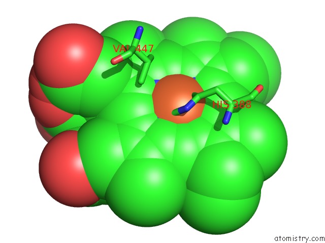
Mono view
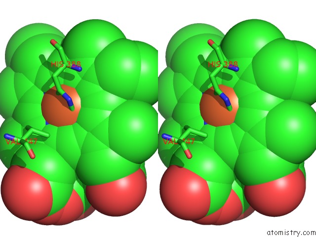
Stereo pair view
Mono view
Stereo pair view
A full contact list of Iron with other atoms in the Fe binding
site number 1 of 2.0 Angstrom Crystal Structure of Ovine Prostaglandin H2 Synthase-1, in Complex with Alpha-Methyl-4-Biphenylacetic Acid within 5.0Å range:
|
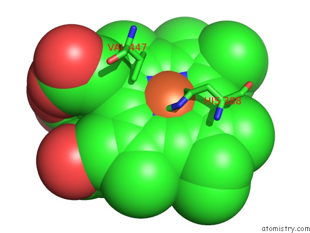
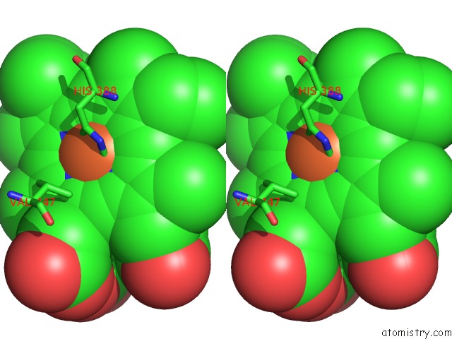
Iron binding site 2 out of 4 in 1q4g
Go back to
Iron binding site 2 out
of 4 in the 2.0 Angstrom Crystal Structure of Ovine Prostaglandin H2 Synthase-1, in Complex with Alpha-Methyl-4-Biphenylacetic Acid

Mono view

Stereo pair view
Mono view
Stereo pair view
A full contact list of Iron with other atoms in the Fe binding
site number 2 of 2.0 Angstrom Crystal Structure of Ovine Prostaglandin H2 Synthase-1, in Complex with Alpha-Methyl-4-Biphenylacetic Acid within 5.0Å range:
|
Iron binding site 3 out of 4 in 1q4g
Go back to
Iron binding site 3 out
of 4 in the 2.0 Angstrom Crystal Structure of Ovine Prostaglandin H2 Synthase-1, in Complex with Alpha-Methyl-4-Biphenylacetic Acid
Mono view
Stereo pair view
Mono view
Stereo pair view
A full contact list of Iron with other atoms in the Fe binding
site number 3 of 2.0 Angstrom Crystal Structure of Ovine Prostaglandin H2 Synthase-1, in Complex with Alpha-Methyl-4-Biphenylacetic Acid within 5.0Å range:
|
Iron binding site 4 out of 4 in 1q4g
Go back to
Iron binding site 4 out
of 4 in the 2.0 Angstrom Crystal Structure of Ovine Prostaglandin H2 Synthase-1, in Complex with Alpha-Methyl-4-Biphenylacetic Acid

Mono view

Stereo pair view
Mono view
Stereo pair view
A full contact list of Iron with other atoms in the Fe binding
site number 4 of 2.0 Angstrom Crystal Structure of Ovine Prostaglandin H2 Synthase-1, in Complex with Alpha-Methyl-4-Biphenylacetic Acid within 5.0Å range:
|
Reference:
K.Gupta,
B.S.Selinsky,
C.J.Kaub,
A.K.Katz,
P.J.Loll.
The 2.0A Resolution Crystal Structure of Prostaglandin H(2) Synthase-1: Structural Insights Into An Unusual Peroxidase J.Mol.Biol. V. 335 503 2004.
ISSN: ISSN 0022-2836
PubMed: 14672659
DOI: 10.1016/J.JMB.2003.10.073
Page generated: Sat Aug 3 13:17:21 2024
ISSN: ISSN 0022-2836
PubMed: 14672659
DOI: 10.1016/J.JMB.2003.10.073
Last articles
Zn in 9J0NZn in 9J0O
Zn in 9J0P
Zn in 9FJX
Zn in 9EKB
Zn in 9C0F
Zn in 9CAH
Zn in 9CH0
Zn in 9CH3
Zn in 9CH1