Iron »
PDB 1qov-1ra5 »
1qvs »
Iron in PDB 1qvs: Crystal Structure of Haemophilus Influenzae H9A Mutant Holo Ferric Ion-Binding Protein A
Protein crystallography data
The structure of Crystal Structure of Haemophilus Influenzae H9A Mutant Holo Ferric Ion-Binding Protein A, PDB code: 1qvs
was solved by
S.R.Shouldice,
R.J.Skene,
D.R.Dougan,
D.E.Mcree,
L.W.Tari,
A.B.Schryvers,
with X-Ray Crystallography technique. A brief refinement statistics is given in the table below:
| Resolution Low / High (Å) | 62.02 / 2.10 |
| Space group | P 21 21 2 |
| Cell size a, b, c (Å), α, β, γ (°) | 105.999, 75.656, 33.774, 90.00, 90.00, 90.00 |
| R / Rfree (%) | 16.8 / 22.6 |
Iron Binding Sites:
The binding sites of Iron atom in the Crystal Structure of Haemophilus Influenzae H9A Mutant Holo Ferric Ion-Binding Protein A
(pdb code 1qvs). This binding sites where shown within
5.0 Angstroms radius around Iron atom.
In total 3 binding sites of Iron where determined in the Crystal Structure of Haemophilus Influenzae H9A Mutant Holo Ferric Ion-Binding Protein A, PDB code: 1qvs:
Jump to Iron binding site number: 1; 2; 3;
In total 3 binding sites of Iron where determined in the Crystal Structure of Haemophilus Influenzae H9A Mutant Holo Ferric Ion-Binding Protein A, PDB code: 1qvs:
Jump to Iron binding site number: 1; 2; 3;
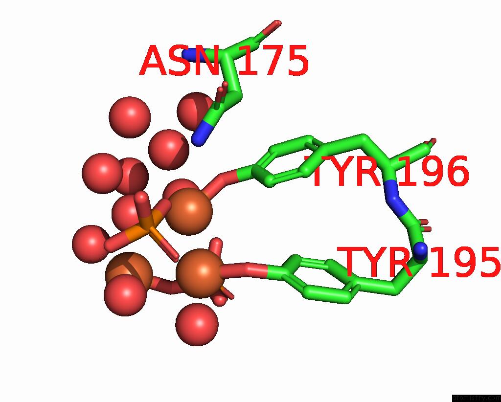
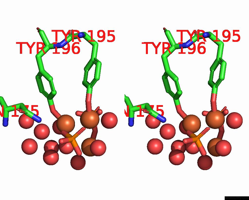
Iron binding site 1 out of 3 in 1qvs
Go back to
Iron binding site 1 out
of 3 in the Crystal Structure of Haemophilus Influenzae H9A Mutant Holo Ferric Ion-Binding Protein A
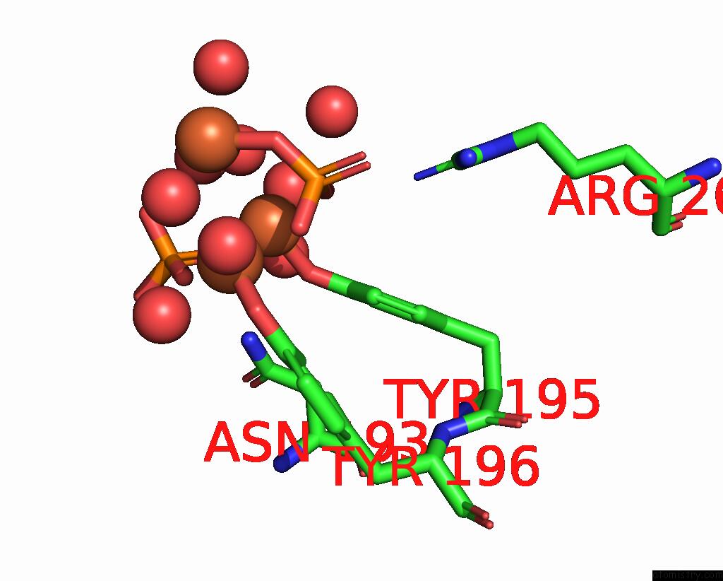
Mono view
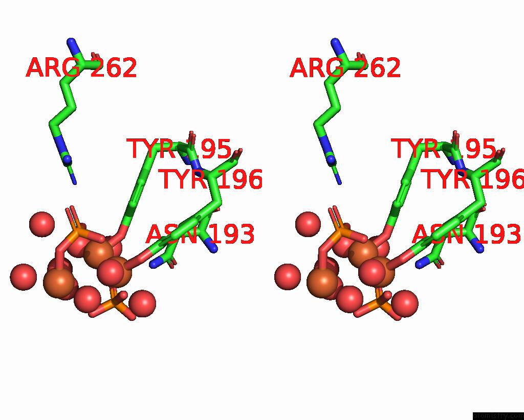
Stereo pair view
Mono view
Stereo pair view
A full contact list of Iron with other atoms in the Fe binding
site number 1 of Crystal Structure of Haemophilus Influenzae H9A Mutant Holo Ferric Ion-Binding Protein A within 5.0Å range:
|
Iron binding site 2 out of 3 in 1qvs
Go back to
Iron binding site 2 out
of 3 in the Crystal Structure of Haemophilus Influenzae H9A Mutant Holo Ferric Ion-Binding Protein A
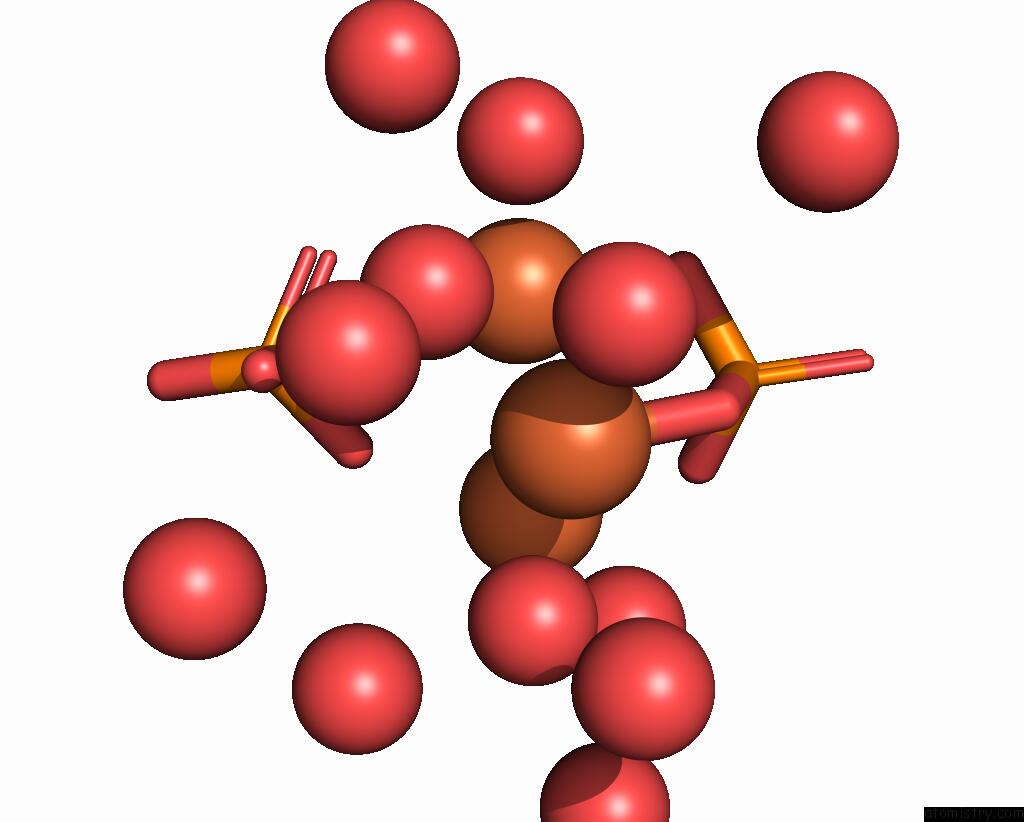
Mono view
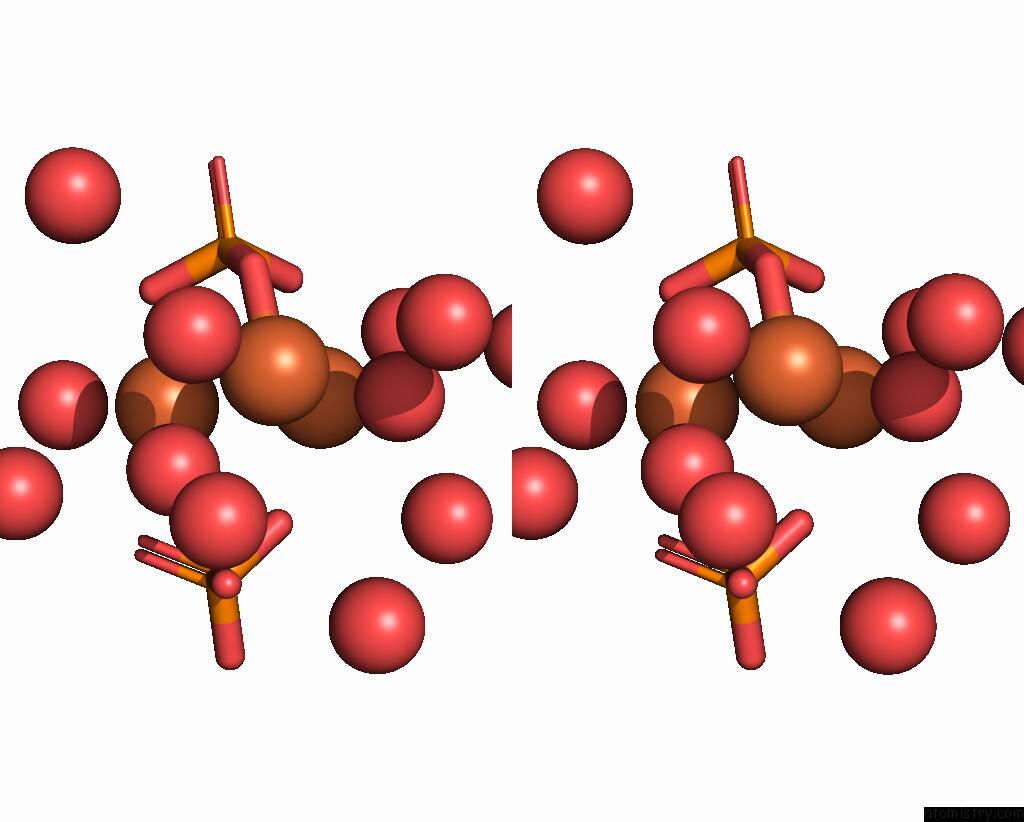
Stereo pair view
Mono view
Stereo pair view
A full contact list of Iron with other atoms in the Fe binding
site number 2 of Crystal Structure of Haemophilus Influenzae H9A Mutant Holo Ferric Ion-Binding Protein A within 5.0Å range:
|
Iron binding site 3 out of 3 in 1qvs
Go back to
Iron binding site 3 out
of 3 in the Crystal Structure of Haemophilus Influenzae H9A Mutant Holo Ferric Ion-Binding Protein A
Mono view
Stereo pair view
Mono view
Stereo pair view
A full contact list of Iron with other atoms in the Fe binding
site number 3 of Crystal Structure of Haemophilus Influenzae H9A Mutant Holo Ferric Ion-Binding Protein A within 5.0Å range:
|
Reference:
S.R.Shouldice,
R.J.Skene,
D.R.Dougan,
D.E.Mcree,
L.W.Tari,
A.B.Schryvers.
Presence of Ferric Hydroxide Clusters in Mutants of Haemophilus Influenzae Ferric Ion-Binding Protein A Biochemistry V. 42 11908 2003.
ISSN: ISSN 0006-2960
PubMed: 14556621
DOI: 10.1021/BI035389S
Page generated: Sat Aug 3 13:52:58 2024
ISSN: ISSN 0006-2960
PubMed: 14556621
DOI: 10.1021/BI035389S
Last articles
Zn in 9J0NZn in 9J0O
Zn in 9J0P
Zn in 9FJX
Zn in 9EKB
Zn in 9C0F
Zn in 9CAH
Zn in 9CH0
Zn in 9CH3
Zn in 9CH1