Iron »
PDB 1qov-1ra5 »
1qyz »
Iron in PDB 1qyz: Characterization of the Malformed, Recombinant Cytochrome RC552
Protein crystallography data
The structure of Characterization of the Malformed, Recombinant Cytochrome RC552, PDB code: 1qyz
was solved by
J.A.Fee,
T.R.Todaro,
E.Luna,
D.Sanders,
L.M.Hunsicker-Wang,
K.M.Patel,
K.L.Bren,
E.Gomez-Moran,
M.G.Hill,
J.Ai,
T.M.Loehr,
W.A.Oertling,
P.A.Williams,
C.D.Stout,
D.Mcree,
A.Pastuszyn,
with X-Ray Crystallography technique. A brief refinement statistics is given in the table below:
| Resolution Low / High (Å) | 30.00 / 1.40 |
| Space group | P 65 |
| Cell size a, b, c (Å), α, β, γ (°) | 87.463, 87.463, 32.209, 90.00, 90.00, 120.00 |
| R / Rfree (%) | 19.7 / 26 |
Iron Binding Sites:
The binding sites of Iron atom in the Characterization of the Malformed, Recombinant Cytochrome RC552
(pdb code 1qyz). This binding sites where shown within
5.0 Angstroms radius around Iron atom.
In total only one binding site of Iron was determined in the Characterization of the Malformed, Recombinant Cytochrome RC552, PDB code: 1qyz:
In total only one binding site of Iron was determined in the Characterization of the Malformed, Recombinant Cytochrome RC552, PDB code: 1qyz:
Iron binding site 1 out of 1 in 1qyz
Go back to
Iron binding site 1 out
of 1 in the Characterization of the Malformed, Recombinant Cytochrome RC552
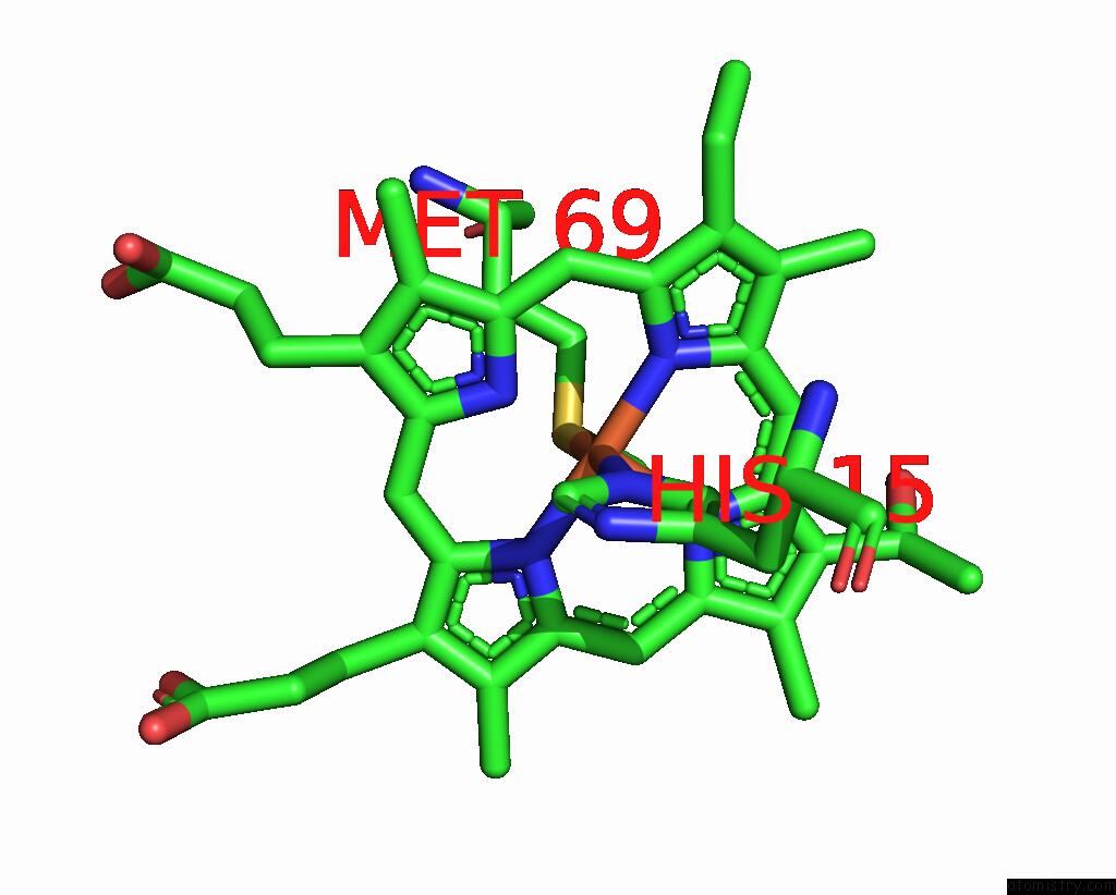
Mono view
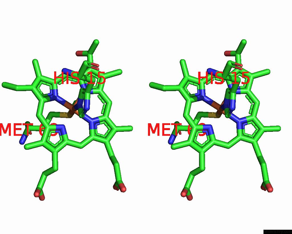
Stereo pair view
Mono view
Stereo pair view
A full contact list of Iron with other atoms in the Fe binding
site number 1 of Characterization of the Malformed, Recombinant Cytochrome RC552 within 5.0Å range:
|
Reference:
J.A.Fee,
T.R.Todaro,
E.Luna,
D.Sanders,
L.M.Hunsicker-Wang,
K.M.Patel,
K.L.Bren,
E.Gomez-Moran,
M.G.Hill,
J.Ai,
T.M.Loehr,
W.A.Oertling,
P.A.Williams,
C.D.Stout,
D.Mcree,
A.Pastuszyn.
Cytochrome RC552, Formed During Expression of the Truncated, Thermus Thermophilus Cytochrome C552 Gene in the Cytoplasm of Escherichia Coli, Reacts Spontaneously to Form Protein-Bound 2-Formyl-4-Vinyl (Spirographis) Heme. Biochemistry V. 43 12162 2004.
ISSN: ISSN 0006-2960
PubMed: 15379555
DOI: 10.1021/BI048968L
Page generated: Sat Aug 3 13:56:49 2024
ISSN: ISSN 0006-2960
PubMed: 15379555
DOI: 10.1021/BI048968L
Last articles
Zn in 9MJ5Zn in 9HNW
Zn in 9G0L
Zn in 9FNE
Zn in 9DZN
Zn in 9E0I
Zn in 9D32
Zn in 9DAK
Zn in 8ZXC
Zn in 8ZUF