Iron »
PDB 1rak-1rsv »
1rfs »
Iron in PDB 1rfs: Rieske Soluble Fragment From Spinach
Protein crystallography data
The structure of Rieske Soluble Fragment From Spinach, PDB code: 1rfs
was solved by
C.J.Carrell,
H.Zhang,
W.A.Cramer,
J.L.Smith,
with X-Ray Crystallography technique. A brief refinement statistics is given in the table below:
| Resolution Low / High (Å) | 20.00 / 1.83 |
| Space group | P 1 |
| Cell size a, b, c (Å), α, β, γ (°) | 29.050, 31.870, 35.790, 95.60, 106.10, 117.30 |
| R / Rfree (%) | 17 / 22 |
Iron Binding Sites:
The binding sites of Iron atom in the Rieske Soluble Fragment From Spinach
(pdb code 1rfs). This binding sites where shown within
5.0 Angstroms radius around Iron atom.
In total 2 binding sites of Iron where determined in the Rieske Soluble Fragment From Spinach, PDB code: 1rfs:
Jump to Iron binding site number: 1; 2;
In total 2 binding sites of Iron where determined in the Rieske Soluble Fragment From Spinach, PDB code: 1rfs:
Jump to Iron binding site number: 1; 2;
Iron binding site 1 out of 2 in 1rfs
Go back to
Iron binding site 1 out
of 2 in the Rieske Soluble Fragment From Spinach

Mono view
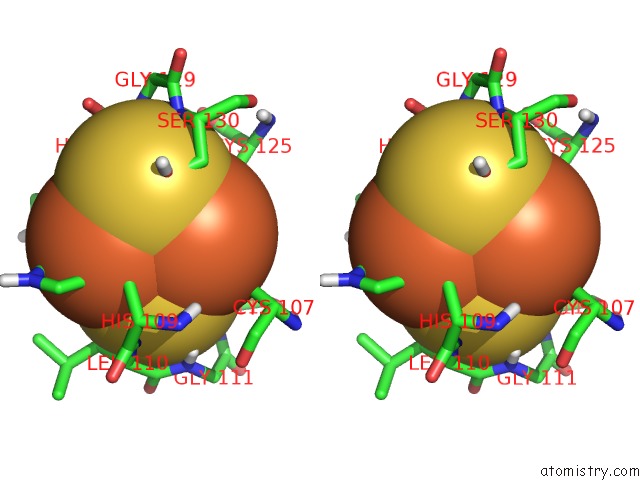
Stereo pair view
Mono view
Stereo pair view
A full contact list of Iron with other atoms in the Fe binding
site number 1 of Rieske Soluble Fragment From Spinach within 5.0Å range:
|
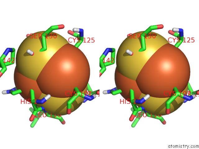
Iron binding site 2 out of 2 in 1rfs
Go back to
Iron binding site 2 out
of 2 in the Rieske Soluble Fragment From Spinach
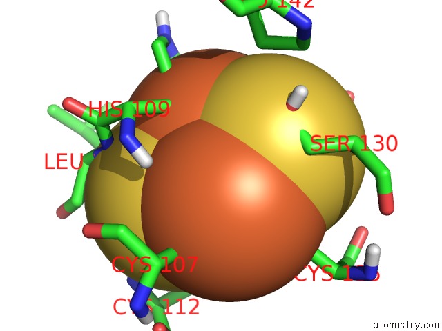
Mono view
Stereo pair view
Mono view
Stereo pair view
A full contact list of Iron with other atoms in the Fe binding
site number 2 of Rieske Soluble Fragment From Spinach within 5.0Å range:
|
Reference:
C.J.Carrell,
H.Zhang,
W.A.Cramer,
J.L.Smith.
Biological Identity and Diversity in Photosynthesis and Respiration: Structure of the Lumen-Side Domain of the Chloroplast Rieske Protein. Structure V. 5 1613 1997.
ISSN: ISSN 0969-2126
PubMed: 9438861
DOI: 10.1016/S0969-2126(97)00309-2
Page generated: Sat Aug 3 14:15:34 2024
ISSN: ISSN 0969-2126
PubMed: 9438861
DOI: 10.1016/S0969-2126(97)00309-2
Last articles
Zn in 9MJ5Zn in 9HNW
Zn in 9G0L
Zn in 9FNE
Zn in 9DZN
Zn in 9E0I
Zn in 9D32
Zn in 9DAK
Zn in 8ZXC
Zn in 8ZUF