Iron »
PDB 1rak-1rsv »
1rom »
Iron in PDB 1rom: Crystal Structure of Nitric Reductase From Denitrifying Fungus Fusarium Oxysporum
Protein crystallography data
The structure of Crystal Structure of Nitric Reductase From Denitrifying Fungus Fusarium Oxysporum, PDB code: 1rom
was solved by
S.-Y.Park,
A.Nakagawa,
with X-Ray Crystallography technique. A brief refinement statistics is given in the table below:
| Resolution Low / High (Å) | 10.00 / 2.00 |
| Space group | P 21 21 21 |
| Cell size a, b, c (Å), α, β, γ (°) | 54.990, 82.660, 87.210, 90.00, 90.00, 90.00 |
| R / Rfree (%) | 19.7 / 26.6 |
Iron Binding Sites:
The binding sites of Iron atom in the Crystal Structure of Nitric Reductase From Denitrifying Fungus Fusarium Oxysporum
(pdb code 1rom). This binding sites where shown within
5.0 Angstroms radius around Iron atom.
In total only one binding site of Iron was determined in the Crystal Structure of Nitric Reductase From Denitrifying Fungus Fusarium Oxysporum, PDB code: 1rom:
In total only one binding site of Iron was determined in the Crystal Structure of Nitric Reductase From Denitrifying Fungus Fusarium Oxysporum, PDB code: 1rom:
Iron binding site 1 out of 1 in 1rom
Go back to
Iron binding site 1 out
of 1 in the Crystal Structure of Nitric Reductase From Denitrifying Fungus Fusarium Oxysporum
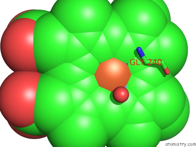
Mono view
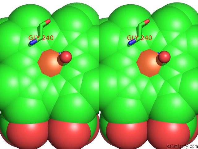
Stereo pair view
Mono view
Stereo pair view
A full contact list of Iron with other atoms in the Fe binding
site number 1 of Crystal Structure of Nitric Reductase From Denitrifying Fungus Fusarium Oxysporum within 5.0Å range:
|
Reference:
S.Y.Park,
H.Shimizu,
S.Adachi,
A.Nakagawa,
I.Tanaka,
K.Nakahara,
H.Shoun,
E.Obayashi,
H.Nakamura,
T.Iizuka,
Y.Shiro.
Crystal Structure of Nitric Oxide Reductase From Denitrifying Fungus Fusarium Oxysporum. Nat.Struct.Biol. V. 4 827 1997.
ISSN: ISSN 1072-8368
PubMed: 9334748
DOI: 10.1038/NSB1097-827
Page generated: Sat Aug 3 14:18:15 2024
ISSN: ISSN 1072-8368
PubMed: 9334748
DOI: 10.1038/NSB1097-827
Last articles
Zn in 9MJ5Zn in 9HNW
Zn in 9G0L
Zn in 9FNE
Zn in 9DZN
Zn in 9E0I
Zn in 9D32
Zn in 9DAK
Zn in 8ZXC
Zn in 8ZUF