Iron »
PDB 1rak-1rsv »
1rse »
Iron in PDB 1rse: Myoglobin (Horse Heart) Mutant with Ser 92 Replaced By Asp (S92D)
Protein crystallography data
The structure of Myoglobin (Horse Heart) Mutant with Ser 92 Replaced By Asp (S92D), PDB code: 1rse
was solved by
D.L.Burk,
G.D.Brayer,
with X-Ray Crystallography technique. A brief refinement statistics is given in the table below:
| Resolution Low / High (Å) | 6.00 / 1.70 |
| Space group | P 1 21 1 |
| Cell size a, b, c (Å), α, β, γ (°) | 64.420, 28.880, 35.970, 90.00, 107.00, 90.00 |
| R / Rfree (%) | 18.8 / n/a |
Iron Binding Sites:
The binding sites of Iron atom in the Myoglobin (Horse Heart) Mutant with Ser 92 Replaced By Asp (S92D)
(pdb code 1rse). This binding sites where shown within
5.0 Angstroms radius around Iron atom.
In total only one binding site of Iron was determined in the Myoglobin (Horse Heart) Mutant with Ser 92 Replaced By Asp (S92D), PDB code: 1rse:
In total only one binding site of Iron was determined in the Myoglobin (Horse Heart) Mutant with Ser 92 Replaced By Asp (S92D), PDB code: 1rse:
Iron binding site 1 out of 1 in 1rse
Go back to
Iron binding site 1 out
of 1 in the Myoglobin (Horse Heart) Mutant with Ser 92 Replaced By Asp (S92D)
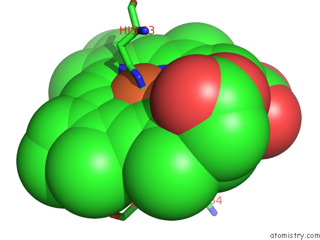
Mono view
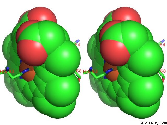
Stereo pair view
Mono view
Stereo pair view
A full contact list of Iron with other atoms in the Fe binding
site number 1 of Myoglobin (Horse Heart) Mutant with Ser 92 Replaced By Asp (S92D) within 5.0Å range:
|
Reference:
E.Lloyd,
D.L.Burk,
J.C.Ferrer,
R.Maurus,
J.Doran,
P.R.Carey,
G.D.Brayer,
A.G.Mauk.
Electrostatic Modification of the Active Site of Myoglobin: Characterization of the Proximal SER92ASP Variant. Biochemistry V. 35 11901 1996.
ISSN: ISSN 0006-2960
PubMed: 8794773
DOI: 10.1021/BI9608976
Page generated: Sat Aug 3 14:24:02 2024
ISSN: ISSN 0006-2960
PubMed: 8794773
DOI: 10.1021/BI9608976
Last articles
Zn in 9MJ5Zn in 9HNW
Zn in 9G0L
Zn in 9FNE
Zn in 9DZN
Zn in 9E0I
Zn in 9D32
Zn in 9DAK
Zn in 8ZXC
Zn in 8ZUF