Iron »
PDB 1rte-1sdk »
1ru3 »
Iron in PDB 1ru3: Crystal Structure of the Monomeric Acetyl-Coa Synthase From Carboxydothermus Hydrogenoformans
Protein crystallography data
The structure of Crystal Structure of the Monomeric Acetyl-Coa Synthase From Carboxydothermus Hydrogenoformans, PDB code: 1ru3
was solved by
V.Svetlitchnyi,
H.Dobbek,
W.Meyer-Klaucke,
T.Meins,
B.Thiele,
P.Rmer,
R.Huber,
O.Meyer,
with X-Ray Crystallography technique. A brief refinement statistics is given in the table below:
| Resolution Low / High (Å) | 20.00 / 2.20 |
| Space group | H 3 2 |
| Cell size a, b, c (Å), α, β, γ (°) | 200.310, 200.310, 169.410, 90.00, 90.00, 120.00 |
| R / Rfree (%) | 23.7 / 27.4 |
Other elements in 1ru3:
The structure of Crystal Structure of the Monomeric Acetyl-Coa Synthase From Carboxydothermus Hydrogenoformans also contains other interesting chemical elements:
| Nickel | (Ni) | 2 atoms |
Iron Binding Sites:
The binding sites of Iron atom in the Crystal Structure of the Monomeric Acetyl-Coa Synthase From Carboxydothermus Hydrogenoformans
(pdb code 1ru3). This binding sites where shown within
5.0 Angstroms radius around Iron atom.
In total 4 binding sites of Iron where determined in the Crystal Structure of the Monomeric Acetyl-Coa Synthase From Carboxydothermus Hydrogenoformans, PDB code: 1ru3:
Jump to Iron binding site number: 1; 2; 3; 4;
In total 4 binding sites of Iron where determined in the Crystal Structure of the Monomeric Acetyl-Coa Synthase From Carboxydothermus Hydrogenoformans, PDB code: 1ru3:
Jump to Iron binding site number: 1; 2; 3; 4;
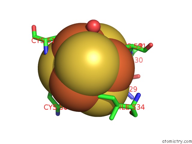
Iron binding site 1 out of 4 in 1ru3
Go back to
Iron binding site 1 out
of 4 in the Crystal Structure of the Monomeric Acetyl-Coa Synthase From Carboxydothermus Hydrogenoformans
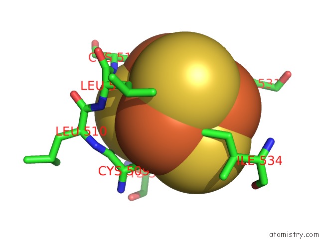
Mono view
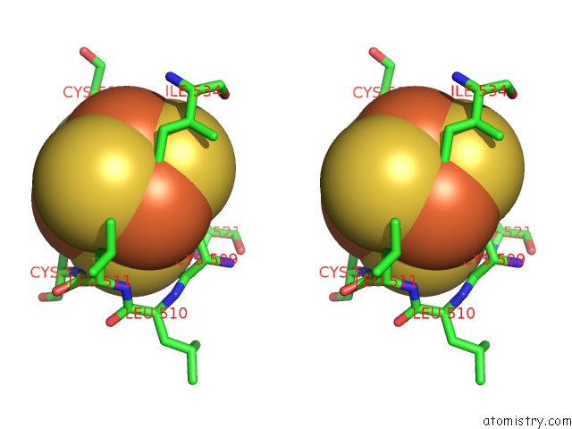
Stereo pair view
Mono view
Stereo pair view
A full contact list of Iron with other atoms in the Fe binding
site number 1 of Crystal Structure of the Monomeric Acetyl-Coa Synthase From Carboxydothermus Hydrogenoformans within 5.0Å range:
|
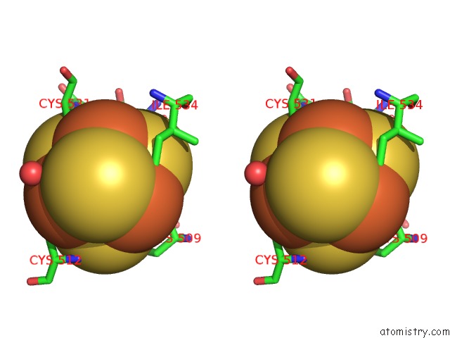
Iron binding site 2 out of 4 in 1ru3
Go back to
Iron binding site 2 out
of 4 in the Crystal Structure of the Monomeric Acetyl-Coa Synthase From Carboxydothermus Hydrogenoformans
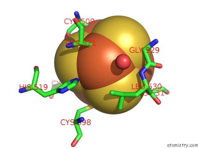
Mono view
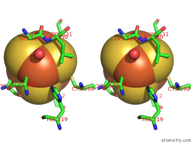
Stereo pair view
Mono view
Stereo pair view
A full contact list of Iron with other atoms in the Fe binding
site number 2 of Crystal Structure of the Monomeric Acetyl-Coa Synthase From Carboxydothermus Hydrogenoformans within 5.0Å range:
|
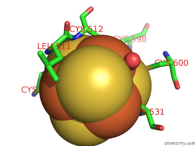
Iron binding site 3 out of 4 in 1ru3
Go back to
Iron binding site 3 out
of 4 in the Crystal Structure of the Monomeric Acetyl-Coa Synthase From Carboxydothermus Hydrogenoformans
Mono view
Stereo pair view
Mono view
Stereo pair view
A full contact list of Iron with other atoms in the Fe binding
site number 3 of Crystal Structure of the Monomeric Acetyl-Coa Synthase From Carboxydothermus Hydrogenoformans within 5.0Å range:
|
Iron binding site 4 out of 4 in 1ru3
Go back to
Iron binding site 4 out
of 4 in the Crystal Structure of the Monomeric Acetyl-Coa Synthase From Carboxydothermus Hydrogenoformans
Mono view
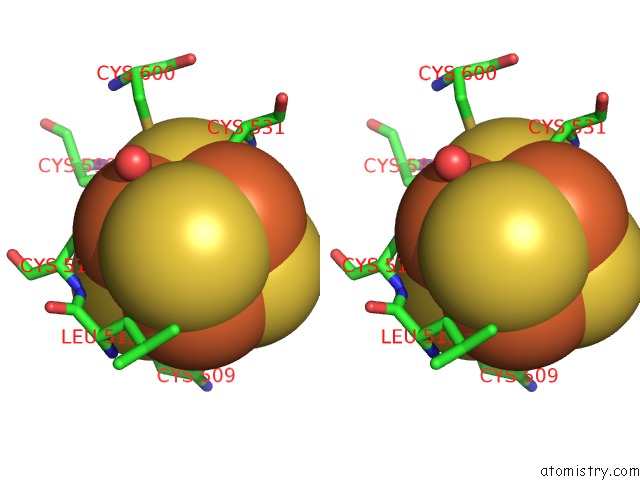
Stereo pair view
Mono view
Stereo pair view
A full contact list of Iron with other atoms in the Fe binding
site number 4 of Crystal Structure of the Monomeric Acetyl-Coa Synthase From Carboxydothermus Hydrogenoformans within 5.0Å range:
|
Reference:
V.Svetlitchnyi,
H.Dobbek,
W.Meyer-Klaucke,
T.Meins,
B.Thiele,
P.Rmer,
R.Huber,
O.Meyer.
A Functional Ni-Ni-[4FE-4S] Cluster in the Monomeric Acetyl-Coa Synthase From Carboxydothermus Hydrogenoformans Proc.Natl.Acad.Sci.Usa V. 101 446 2004.
ISSN: ISSN 0027-8424
PubMed: 14699043
DOI: 10.1073/PNAS.0304262101
Page generated: Sat Aug 3 14:30:04 2024
ISSN: ISSN 0027-8424
PubMed: 14699043
DOI: 10.1073/PNAS.0304262101
Last articles
Zn in 9MJ5Zn in 9HNW
Zn in 9G0L
Zn in 9FNE
Zn in 9DZN
Zn in 9E0I
Zn in 9D32
Zn in 9DAK
Zn in 8ZXC
Zn in 8ZUF