Iron »
PDB 1rte-1sdk »
1ry5 »
Iron in PDB 1ry5: Photosynthetic Reaction Center Mutant From Rhodobacter Sphaeroides with Asp L213 Replaced with Asn
Protein crystallography data
The structure of Photosynthetic Reaction Center Mutant From Rhodobacter Sphaeroides with Asp L213 Replaced with Asn, PDB code: 1ry5
was solved by
Q.Xu,
H.L.Axelrod,
E.C.Abresch,
M.L.Paddock,
M.Y.Okamura,
G.Feher,
with X-Ray Crystallography technique. A brief refinement statistics is given in the table below:
| Resolution Low / High (Å) | 39.23 / 2.10 |
| Space group | P 31 2 1 |
| Cell size a, b, c (Å), α, β, γ (°) | 139.071, 139.071, 184.599, 90.00, 90.00, 120.00 |
| R / Rfree (%) | 21.1 / 22.6 |
Other elements in 1ry5:
The structure of Photosynthetic Reaction Center Mutant From Rhodobacter Sphaeroides with Asp L213 Replaced with Asn also contains other interesting chemical elements:
| Magnesium | (Mg) | 4 atoms |
| Potassium | (K) | 1 atom |
Iron Binding Sites:
The binding sites of Iron atom in the Photosynthetic Reaction Center Mutant From Rhodobacter Sphaeroides with Asp L213 Replaced with Asn
(pdb code 1ry5). This binding sites where shown within
5.0 Angstroms radius around Iron atom.
In total only one binding site of Iron was determined in the Photosynthetic Reaction Center Mutant From Rhodobacter Sphaeroides with Asp L213 Replaced with Asn, PDB code: 1ry5:
In total only one binding site of Iron was determined in the Photosynthetic Reaction Center Mutant From Rhodobacter Sphaeroides with Asp L213 Replaced with Asn, PDB code: 1ry5:
Iron binding site 1 out of 1 in 1ry5
Go back to
Iron binding site 1 out
of 1 in the Photosynthetic Reaction Center Mutant From Rhodobacter Sphaeroides with Asp L213 Replaced with Asn
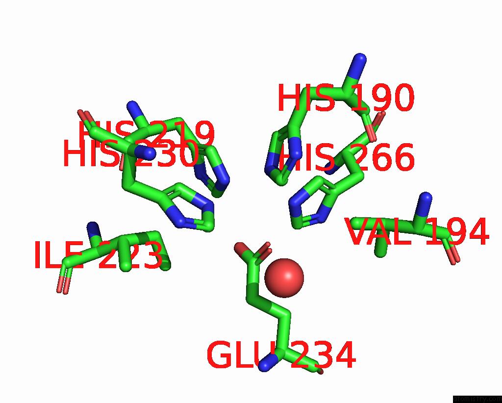
Mono view
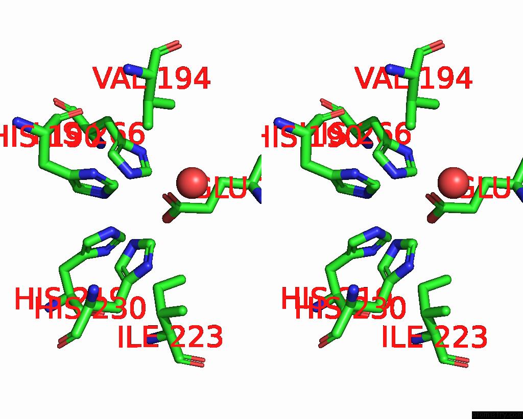
Stereo pair view
Mono view
Stereo pair view
A full contact list of Iron with other atoms in the Fe binding
site number 1 of Photosynthetic Reaction Center Mutant From Rhodobacter Sphaeroides with Asp L213 Replaced with Asn within 5.0Å range:
|
Reference:
Q.Xu,
H.L.Axelrod,
E.C.Abresch,
M.L.Paddock,
M.Y.Okamura,
G.Feher.
X-Ray Structure Determination of Three Mutants of the Bacterial Photosynthetic Reaction Centers From Rb. Sphaeroides; Altered Proton Transfer Pathways. Structure V. 12 703 2004.
ISSN: ISSN 0969-2126
PubMed: 15062092
DOI: 10.1016/J.STR.2004.03.001
Page generated: Sat Aug 3 14:30:05 2024
ISSN: ISSN 0969-2126
PubMed: 15062092
DOI: 10.1016/J.STR.2004.03.001
Last articles
Zn in 9MJ5Zn in 9HNW
Zn in 9G0L
Zn in 9FNE
Zn in 9DZN
Zn in 9E0I
Zn in 9D32
Zn in 9DAK
Zn in 8ZXC
Zn in 8ZUF