Iron »
PDB 1su6-1tfd »
1suv »
Iron in PDB 1suv: Structure of Human Transferrin Receptor-Transferrin Complex
Iron Binding Sites:
The binding sites of Iron atom in the Structure of Human Transferrin Receptor-Transferrin Complex
(pdb code 1suv). This binding sites where shown within
5.0 Angstroms radius around Iron atom.
In total 4 binding sites of Iron where determined in the Structure of Human Transferrin Receptor-Transferrin Complex, PDB code: 1suv:
Jump to Iron binding site number: 1; 2; 3; 4;
In total 4 binding sites of Iron where determined in the Structure of Human Transferrin Receptor-Transferrin Complex, PDB code: 1suv:
Jump to Iron binding site number: 1; 2; 3; 4;
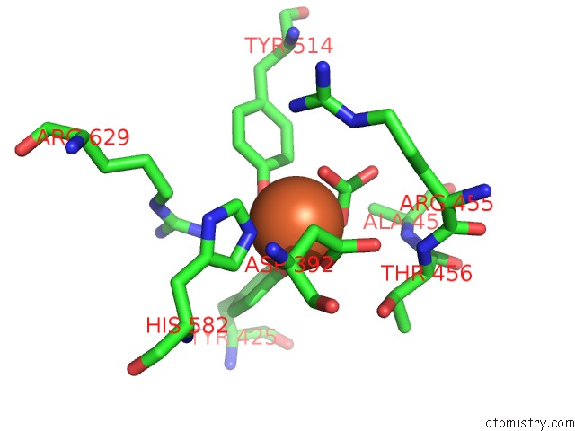
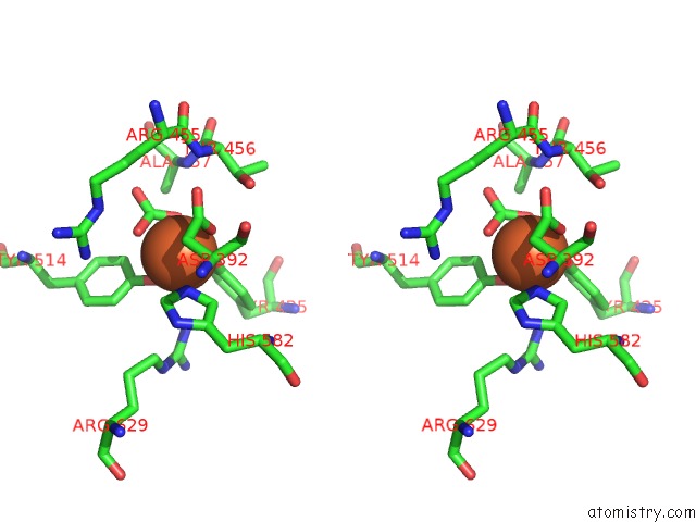
Iron binding site 1 out of 4 in 1suv
Go back to
Iron binding site 1 out
of 4 in the Structure of Human Transferrin Receptor-Transferrin Complex
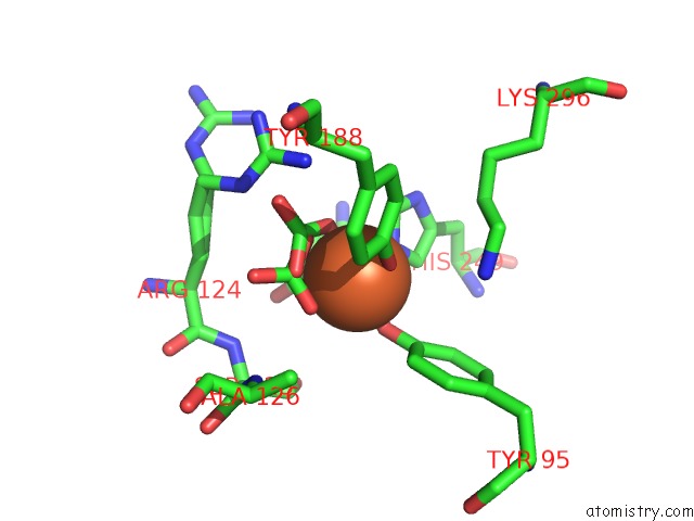
Mono view
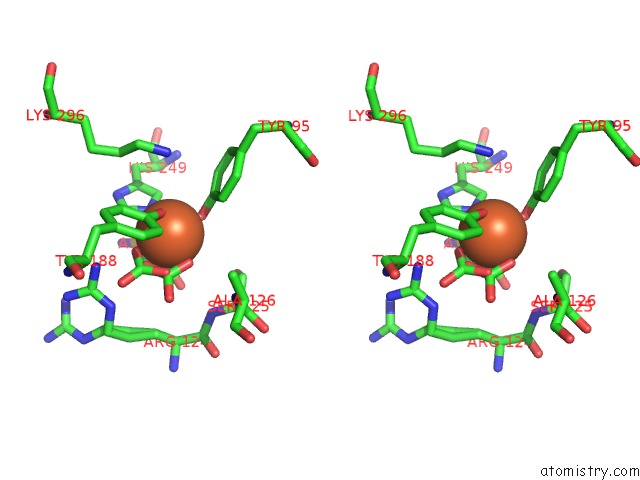
Stereo pair view
Mono view
Stereo pair view
A full contact list of Iron with other atoms in the Fe binding
site number 1 of Structure of Human Transferrin Receptor-Transferrin Complex within 5.0Å range:
|
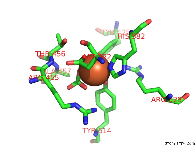
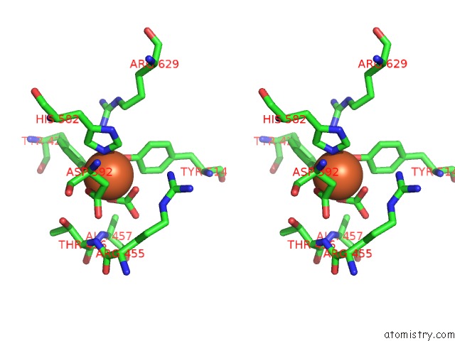
Iron binding site 2 out of 4 in 1suv
Go back to
Iron binding site 2 out
of 4 in the Structure of Human Transferrin Receptor-Transferrin Complex
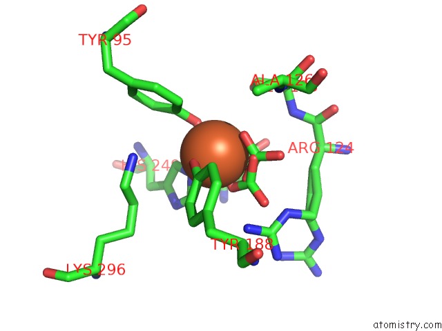
Mono view
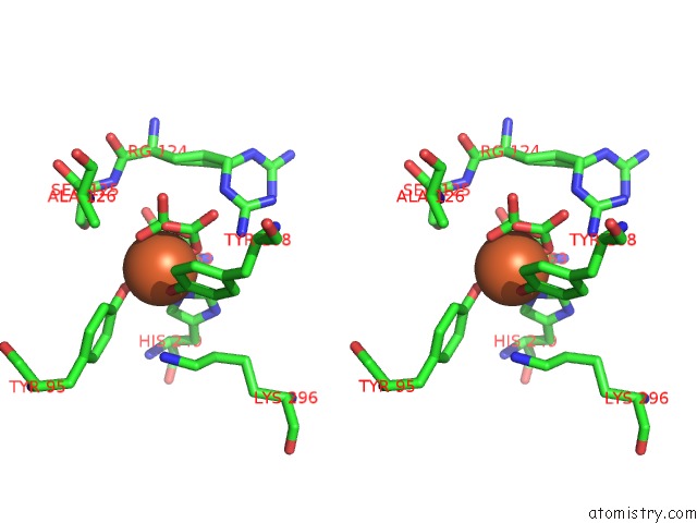
Stereo pair view
Mono view
Stereo pair view
A full contact list of Iron with other atoms in the Fe binding
site number 2 of Structure of Human Transferrin Receptor-Transferrin Complex within 5.0Å range:
|
Iron binding site 3 out of 4 in 1suv
Go back to
Iron binding site 3 out
of 4 in the Structure of Human Transferrin Receptor-Transferrin Complex
Mono view
Stereo pair view
Mono view
Stereo pair view
A full contact list of Iron with other atoms in the Fe binding
site number 3 of Structure of Human Transferrin Receptor-Transferrin Complex within 5.0Å range:
|
Iron binding site 4 out of 4 in 1suv
Go back to
Iron binding site 4 out
of 4 in the Structure of Human Transferrin Receptor-Transferrin Complex
Mono view
Stereo pair view
Mono view
Stereo pair view
A full contact list of Iron with other atoms in the Fe binding
site number 4 of Structure of Human Transferrin Receptor-Transferrin Complex within 5.0Å range:
|
Reference:
Y.Cheng,
O.Zak,
P.Aisen,
S.C.Harrison,
T.Walz.
Structure of the Human Transferrin Receptor-Transferrin Complex Cell(Cambridge,Mass.) V. 116 565 2004.
ISSN: ISSN 0092-8674
PubMed: 14980223
DOI: 10.1016/S0092-8674(04)00130-8
Page generated: Sat Aug 3 15:03:16 2024
ISSN: ISSN 0092-8674
PubMed: 14980223
DOI: 10.1016/S0092-8674(04)00130-8
Last articles
Zn in 9MJ5Zn in 9HNW
Zn in 9G0L
Zn in 9FNE
Zn in 9DZN
Zn in 9E0I
Zn in 9D32
Zn in 9DAK
Zn in 8ZXC
Zn in 8ZUF