Iron »
PDB 1su6-1tfd »
1sxx »
Iron in PDB 1sxx: 1.0 A Crystal Structure of D129A/L130A Mutant of Nitrophorin 4 Complexed with Nitric Oxide
Protein crystallography data
The structure of 1.0 A Crystal Structure of D129A/L130A Mutant of Nitrophorin 4 Complexed with Nitric Oxide, PDB code: 1sxx
was solved by
E.M.Maes,
A.Weichsel,
J.F.Andersen,
D.Shepley,
W.R.Montfort,
with X-Ray Crystallography technique. A brief refinement statistics is given in the table below:
| Resolution Low / High (Å) | 30.57 / 1.01 |
| Space group | C 1 2 1 |
| Cell size a, b, c (Å), α, β, γ (°) | 70.219, 42.751, 53.055, 90.00, 94.30, 90.00 |
| R / Rfree (%) | 14.1 / 15.5 |
Iron Binding Sites:
The binding sites of Iron atom in the 1.0 A Crystal Structure of D129A/L130A Mutant of Nitrophorin 4 Complexed with Nitric Oxide
(pdb code 1sxx). This binding sites where shown within
5.0 Angstroms radius around Iron atom.
In total only one binding site of Iron was determined in the 1.0 A Crystal Structure of D129A/L130A Mutant of Nitrophorin 4 Complexed with Nitric Oxide, PDB code: 1sxx:
In total only one binding site of Iron was determined in the 1.0 A Crystal Structure of D129A/L130A Mutant of Nitrophorin 4 Complexed with Nitric Oxide, PDB code: 1sxx:
Iron binding site 1 out of 1 in 1sxx
Go back to
Iron binding site 1 out
of 1 in the 1.0 A Crystal Structure of D129A/L130A Mutant of Nitrophorin 4 Complexed with Nitric Oxide
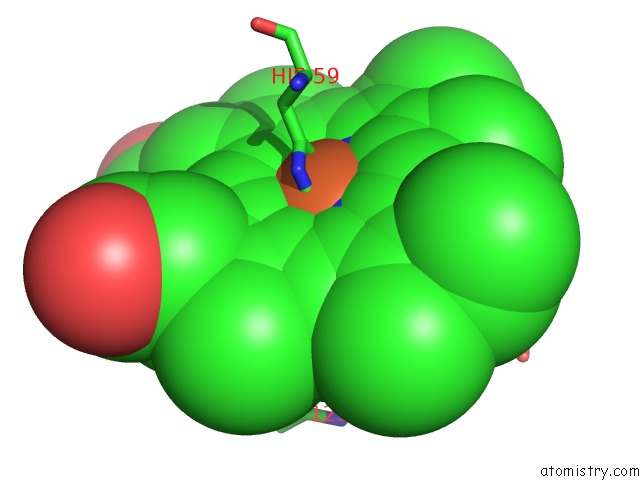
Mono view
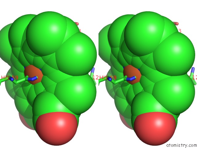
Stereo pair view
Mono view
Stereo pair view
A full contact list of Iron with other atoms in the Fe binding
site number 1 of 1.0 A Crystal Structure of D129A/L130A Mutant of Nitrophorin 4 Complexed with Nitric Oxide within 5.0Å range:
|
Reference:
E.M.Maes,
A.Weichsel,
J.F.Andersen,
D.Shepley,
W.R.Montfort.
Role of Binding Site Loops in Controlling Nitric Oxide Release: Structure and Kinetics of Mutant Forms of Nitrophorin 4 Biochemistry V. 43 6679 2004.
ISSN: ISSN 0006-2960
PubMed: 15157102
DOI: 10.1021/BI049748A
Page generated: Sat Aug 3 15:04:25 2024
ISSN: ISSN 0006-2960
PubMed: 15157102
DOI: 10.1021/BI049748A
Last articles
F in 4QRCF in 4QJW
F in 4QQE
F in 4QR6
F in 4QPF
F in 4QJO
F in 4QMV
F in 4QMZ
F in 4QL1
F in 4QMU