Iron »
PDB 1su6-1tfd »
1t1n »
Iron in PDB 1t1n: Crystal Structure of Carbonmonoxy Hemoglobin
Protein crystallography data
The structure of Crystal Structure of Carbonmonoxy Hemoglobin, PDB code: 1t1n
was solved by
L.Mazzarella,
L.Vitagliano,
C.Savino,
A.Zagari,
with X-Ray Crystallography technique. A brief refinement statistics is given in the table below:
| Resolution Low / High (Å) | 16.00 / 2.20 |
| Space group | C 1 2 1 |
| Cell size a, b, c (Å), α, β, γ (°) | 91.170, 88.060, 55.250, 90.00, 97.65, 90.00 |
| R / Rfree (%) | 19.2 / n/a |
Iron Binding Sites:
The binding sites of Iron atom in the Crystal Structure of Carbonmonoxy Hemoglobin
(pdb code 1t1n). This binding sites where shown within
5.0 Angstroms radius around Iron atom.
In total 2 binding sites of Iron where determined in the Crystal Structure of Carbonmonoxy Hemoglobin, PDB code: 1t1n:
Jump to Iron binding site number: 1; 2;
In total 2 binding sites of Iron where determined in the Crystal Structure of Carbonmonoxy Hemoglobin, PDB code: 1t1n:
Jump to Iron binding site number: 1; 2;
Iron binding site 1 out of 2 in 1t1n
Go back to
Iron binding site 1 out
of 2 in the Crystal Structure of Carbonmonoxy Hemoglobin
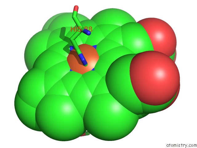
Mono view
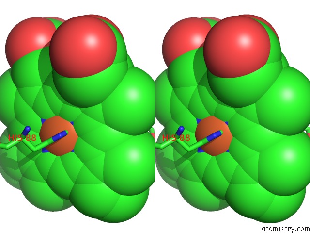
Stereo pair view
Mono view
Stereo pair view
A full contact list of Iron with other atoms in the Fe binding
site number 1 of Crystal Structure of Carbonmonoxy Hemoglobin within 5.0Å range:
|
Iron binding site 2 out of 2 in 1t1n
Go back to
Iron binding site 2 out
of 2 in the Crystal Structure of Carbonmonoxy Hemoglobin
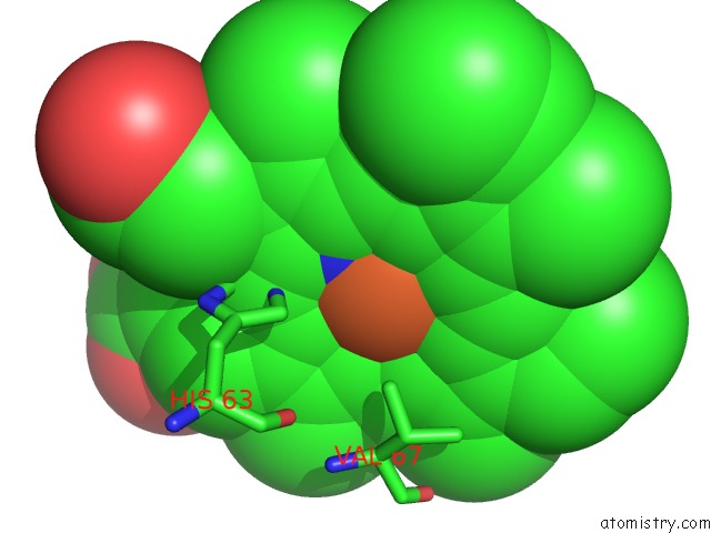
Mono view

Stereo pair view
Mono view
Stereo pair view
A full contact list of Iron with other atoms in the Fe binding
site number 2 of Crystal Structure of Carbonmonoxy Hemoglobin within 5.0Å range:
|
Reference:
L.Mazzarella,
R.D'avino,
G.Di Prisco,
C.Savino,
L.Vitagliano,
P.C.E.Moody,
A.Zagari.
Crystal Structure of Trematomus Newnesi Haemoglobin Re-Opens the Root Effect Question. J.Mol.Biol. V. 287 897 1999.
ISSN: ISSN 0022-2836
PubMed: 10222199
DOI: 10.1006/JMBI.1999.2632
Page generated: Sat Aug 3 15:07:48 2024
ISSN: ISSN 0022-2836
PubMed: 10222199
DOI: 10.1006/JMBI.1999.2632
Last articles
Zn in 9J0NZn in 9J0O
Zn in 9J0P
Zn in 9FJX
Zn in 9EKB
Zn in 9C0F
Zn in 9CAH
Zn in 9CH0
Zn in 9CH3
Zn in 9CH1