Iron »
PDB 1su6-1tfd »
1t3q »
Iron in PDB 1t3q: Crystal Structure of Quinoline 2-Oxidoreductase From Pseudomonas Putida 86
Enzymatic activity of Crystal Structure of Quinoline 2-Oxidoreductase From Pseudomonas Putida 86
All present enzymatic activity of Crystal Structure of Quinoline 2-Oxidoreductase From Pseudomonas Putida 86:
1.3.99.17;
1.3.99.17;
Protein crystallography data
The structure of Crystal Structure of Quinoline 2-Oxidoreductase From Pseudomonas Putida 86, PDB code: 1t3q
was solved by
I.Bonin,
B.M.Martins,
V.Purvanov,
S.Fetzner,
R.Huber,
H.Dobbek,
with X-Ray Crystallography technique. A brief refinement statistics is given in the table below:
| Resolution Low / High (Å) | 19.29 / 1.80 |
| Space group | C 1 2 1 |
| Cell size a, b, c (Å), α, β, γ (°) | 278.320, 72.100, 202.650, 90.00, 127.98, 90.00 |
| R / Rfree (%) | 18.6 / 20.7 |
Other elements in 1t3q:
The structure of Crystal Structure of Quinoline 2-Oxidoreductase From Pseudomonas Putida 86 also contains other interesting chemical elements:
| Molybdenum | (Mo) | 2 atoms |
Iron Binding Sites:
The binding sites of Iron atom in the Crystal Structure of Quinoline 2-Oxidoreductase From Pseudomonas Putida 86
(pdb code 1t3q). This binding sites where shown within
5.0 Angstroms radius around Iron atom.
In total 8 binding sites of Iron where determined in the Crystal Structure of Quinoline 2-Oxidoreductase From Pseudomonas Putida 86, PDB code: 1t3q:
Jump to Iron binding site number: 1; 2; 3; 4; 5; 6; 7; 8;
In total 8 binding sites of Iron where determined in the Crystal Structure of Quinoline 2-Oxidoreductase From Pseudomonas Putida 86, PDB code: 1t3q:
Jump to Iron binding site number: 1; 2; 3; 4; 5; 6; 7; 8;
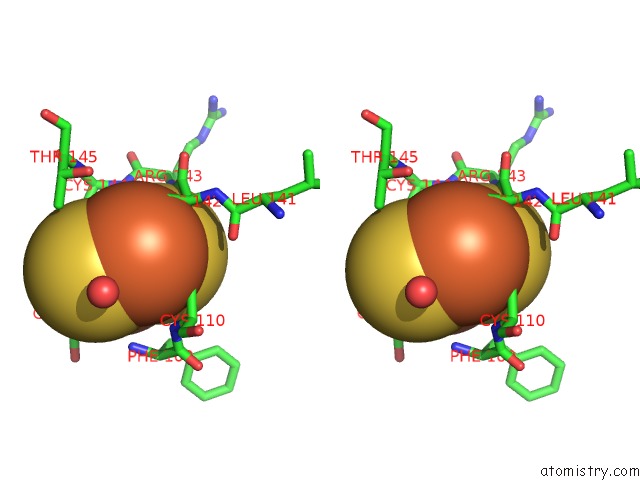
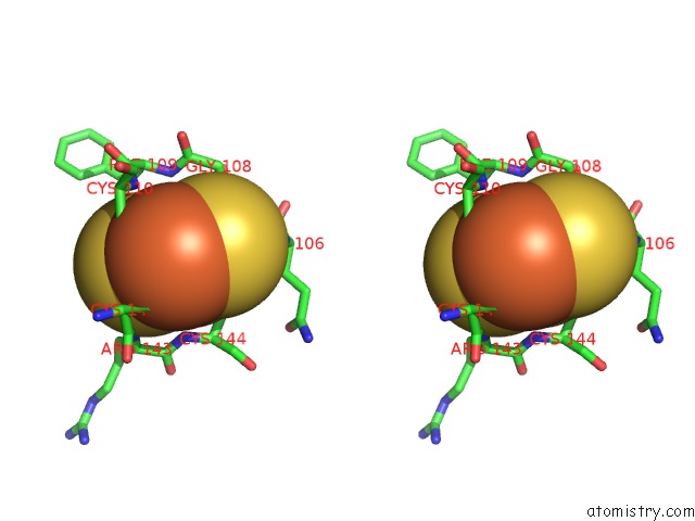
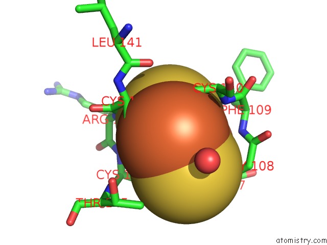
Iron binding site 1 out of 8 in 1t3q
Go back to
Iron binding site 1 out
of 8 in the Crystal Structure of Quinoline 2-Oxidoreductase From Pseudomonas Putida 86
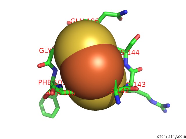
Mono view

Stereo pair view
Mono view
Stereo pair view
A full contact list of Iron with other atoms in the Fe binding
site number 1 of Crystal Structure of Quinoline 2-Oxidoreductase From Pseudomonas Putida 86 within 5.0Å range:
|
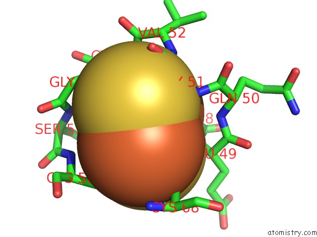
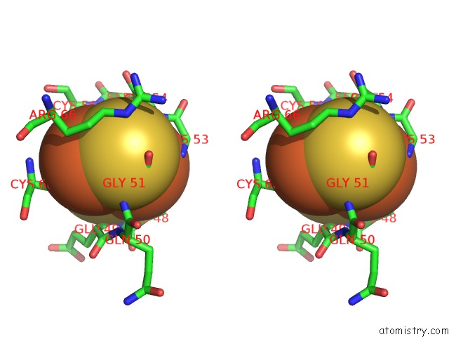
Iron binding site 2 out of 8 in 1t3q
Go back to
Iron binding site 2 out
of 8 in the Crystal Structure of Quinoline 2-Oxidoreductase From Pseudomonas Putida 86

Mono view
Stereo pair view
Mono view
Stereo pair view
A full contact list of Iron with other atoms in the Fe binding
site number 2 of Crystal Structure of Quinoline 2-Oxidoreductase From Pseudomonas Putida 86 within 5.0Å range:
|
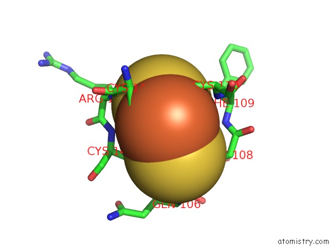
Iron binding site 3 out of 8 in 1t3q
Go back to
Iron binding site 3 out
of 8 in the Crystal Structure of Quinoline 2-Oxidoreductase From Pseudomonas Putida 86
Mono view

Stereo pair view
Mono view
Stereo pair view
A full contact list of Iron with other atoms in the Fe binding
site number 3 of Crystal Structure of Quinoline 2-Oxidoreductase From Pseudomonas Putida 86 within 5.0Å range:
|
Iron binding site 4 out of 8 in 1t3q
Go back to
Iron binding site 4 out
of 8 in the Crystal Structure of Quinoline 2-Oxidoreductase From Pseudomonas Putida 86

Mono view

Stereo pair view
Mono view
Stereo pair view
A full contact list of Iron with other atoms in the Fe binding
site number 4 of Crystal Structure of Quinoline 2-Oxidoreductase From Pseudomonas Putida 86 within 5.0Å range:
|
Iron binding site 5 out of 8 in 1t3q
Go back to
Iron binding site 5 out
of 8 in the Crystal Structure of Quinoline 2-Oxidoreductase From Pseudomonas Putida 86
Mono view
Stereo pair view
Mono view
Stereo pair view
A full contact list of Iron with other atoms in the Fe binding
site number 5 of Crystal Structure of Quinoline 2-Oxidoreductase From Pseudomonas Putida 86 within 5.0Å range:
|
Iron binding site 6 out of 8 in 1t3q
Go back to
Iron binding site 6 out
of 8 in the Crystal Structure of Quinoline 2-Oxidoreductase From Pseudomonas Putida 86
Mono view

Stereo pair view
Mono view
Stereo pair view
A full contact list of Iron with other atoms in the Fe binding
site number 6 of Crystal Structure of Quinoline 2-Oxidoreductase From Pseudomonas Putida 86 within 5.0Å range:
|
Iron binding site 7 out of 8 in 1t3q
Go back to
Iron binding site 7 out
of 8 in the Crystal Structure of Quinoline 2-Oxidoreductase From Pseudomonas Putida 86

Mono view

Stereo pair view
Mono view
Stereo pair view
A full contact list of Iron with other atoms in the Fe binding
site number 7 of Crystal Structure of Quinoline 2-Oxidoreductase From Pseudomonas Putida 86 within 5.0Å range:
|
Iron binding site 8 out of 8 in 1t3q
Go back to
Iron binding site 8 out
of 8 in the Crystal Structure of Quinoline 2-Oxidoreductase From Pseudomonas Putida 86

Mono view
Stereo pair view
Mono view
Stereo pair view
A full contact list of Iron with other atoms in the Fe binding
site number 8 of Crystal Structure of Quinoline 2-Oxidoreductase From Pseudomonas Putida 86 within 5.0Å range:
|
Reference:
I.Bonin,
B.M.Martins,
V.Purvanov,
S.Fetzner,
R.Huber,
H.Dobbek.
Active Site Geometry and Substrate Recognition of the Molybdenum Hydroxylase Quinoline 2-Oxidoreductase. Structure V. 12 1425 2004.
ISSN: ISSN 0969-2126
PubMed: 15296736
DOI: 10.1016/J.STR.2004.05.014
Page generated: Wed Jul 16 20:57:57 2025
ISSN: ISSN 0969-2126
PubMed: 15296736
DOI: 10.1016/J.STR.2004.05.014
Last articles
Fe in 2YXOFe in 2YRS
Fe in 2YXC
Fe in 2YNM
Fe in 2YVJ
Fe in 2YP1
Fe in 2YU2
Fe in 2YU1
Fe in 2YQB
Fe in 2YOO