Iron »
PDB 1su6-1tfd »
1tdw »
Iron in PDB 1tdw: Crystal Structure of Double Truncated Human Phenylalanine Hydroxylase BH4-Responsive Pku Mutant A313T.
Enzymatic activity of Crystal Structure of Double Truncated Human Phenylalanine Hydroxylase BH4-Responsive Pku Mutant A313T.
All present enzymatic activity of Crystal Structure of Double Truncated Human Phenylalanine Hydroxylase BH4-Responsive Pku Mutant A313T.:
1.14.16.1;
1.14.16.1;
Protein crystallography data
The structure of Crystal Structure of Double Truncated Human Phenylalanine Hydroxylase BH4-Responsive Pku Mutant A313T., PDB code: 1tdw
was solved by
H.Erlandsen,
A.L.Pey,
A.Gamez,
B.Perez,
L.R.Desviat,
C.Aguado,
R.Koch,
S.Surendran,
S.Tyring,
R.Matalon,
C.R.Scriver,
M.Ugarte,
A.Martinez,
R.C.Stevens,
with X-Ray Crystallography technique. A brief refinement statistics is given in the table below:
| Resolution Low / High (Å) | 19.93 / 2.10 |
| Space group | C 2 2 21 |
| Cell size a, b, c (Å), α, β, γ (°) | 66.411, 108.138, 124.620, 90.00, 90.00, 90.00 |
| R / Rfree (%) | 20.6 / 23.7 |
Iron Binding Sites:
The binding sites of Iron atom in the Crystal Structure of Double Truncated Human Phenylalanine Hydroxylase BH4-Responsive Pku Mutant A313T.
(pdb code 1tdw). This binding sites where shown within
5.0 Angstroms radius around Iron atom.
In total only one binding site of Iron was determined in the Crystal Structure of Double Truncated Human Phenylalanine Hydroxylase BH4-Responsive Pku Mutant A313T., PDB code: 1tdw:
In total only one binding site of Iron was determined in the Crystal Structure of Double Truncated Human Phenylalanine Hydroxylase BH4-Responsive Pku Mutant A313T., PDB code: 1tdw:
Iron binding site 1 out of 1 in 1tdw
Go back to
Iron binding site 1 out
of 1 in the Crystal Structure of Double Truncated Human Phenylalanine Hydroxylase BH4-Responsive Pku Mutant A313T.
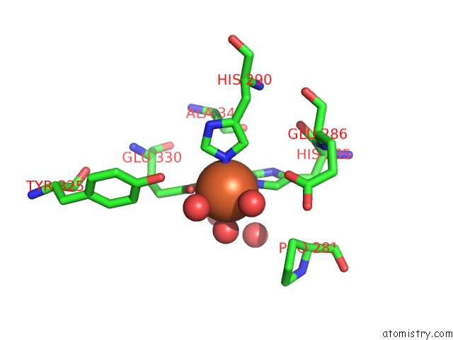
Mono view
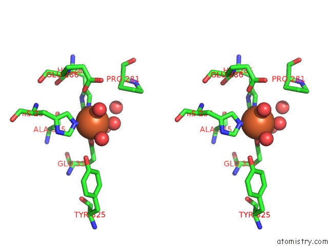
Stereo pair view
Mono view
Stereo pair view
A full contact list of Iron with other atoms in the Fe binding
site number 1 of Crystal Structure of Double Truncated Human Phenylalanine Hydroxylase BH4-Responsive Pku Mutant A313T. within 5.0Å range:
|
Reference:
H.Erlandsen,
A.L.Pey,
A.Gamez,
B.Perez,
L.R.Desviat,
C.Aguado,
R.Koch,
S.Surendran,
S.Tyring,
R.Matalon,
C.R.Scriver,
M.Ugarte,
A.Martinez,
R.C.Stevens.
Correction of Kinetic and Stability Defects By Tetrahydrobiopterin in Phenylketonuria Patients with Certain Phenylalanine Hydroxylase Mutations. Proc.Natl.Acad.Sci.Usa V. 101 16903 2004.
ISSN: ISSN 0027-8424
PubMed: 15557004
DOI: 10.1073/PNAS.0407256101
Page generated: Sat Aug 3 15:13:57 2024
ISSN: ISSN 0027-8424
PubMed: 15557004
DOI: 10.1073/PNAS.0407256101
Last articles
Zn in 9MJ5Zn in 9HNW
Zn in 9G0L
Zn in 9FNE
Zn in 9DZN
Zn in 9E0I
Zn in 9D32
Zn in 9DAK
Zn in 8ZXC
Zn in 8ZUF