Iron »
PDB 1tfz-1ubh »
1toh »
Iron in PDB 1toh: Tyrosine Hydroxylase Catalytic and Tetramerization Domains From Rat
Enzymatic activity of Tyrosine Hydroxylase Catalytic and Tetramerization Domains From Rat
All present enzymatic activity of Tyrosine Hydroxylase Catalytic and Tetramerization Domains From Rat:
1.14.16.2;
1.14.16.2;
Protein crystallography data
The structure of Tyrosine Hydroxylase Catalytic and Tetramerization Domains From Rat, PDB code: 1toh
was solved by
K.E.Goodwill,
C.Sabatier,
R.C.Stevens,
with X-Ray Crystallography technique. A brief refinement statistics is given in the table below:
| Resolution Low / High (Å) | 37.00 / 2.30 |
| Space group | F 2 2 2 |
| Cell size a, b, c (Å), α, β, γ (°) | 189.323, 148.335, 57.975, 90.00, 90.00, 90.00 |
| R / Rfree (%) | 21.1 / 26.8 |
Iron Binding Sites:
The binding sites of Iron atom in the Tyrosine Hydroxylase Catalytic and Tetramerization Domains From Rat
(pdb code 1toh). This binding sites where shown within
5.0 Angstroms radius around Iron atom.
In total only one binding site of Iron was determined in the Tyrosine Hydroxylase Catalytic and Tetramerization Domains From Rat, PDB code: 1toh:
In total only one binding site of Iron was determined in the Tyrosine Hydroxylase Catalytic and Tetramerization Domains From Rat, PDB code: 1toh:
Iron binding site 1 out of 1 in 1toh
Go back to
Iron binding site 1 out
of 1 in the Tyrosine Hydroxylase Catalytic and Tetramerization Domains From Rat
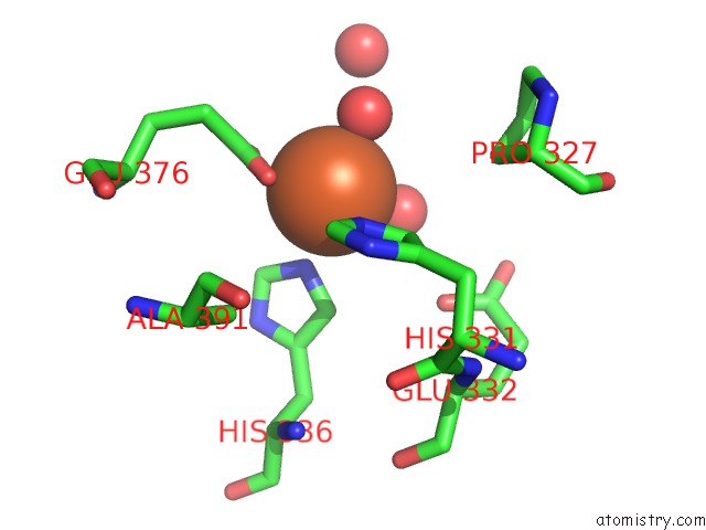
Mono view
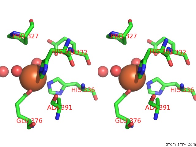
Stereo pair view
Mono view
Stereo pair view
A full contact list of Iron with other atoms in the Fe binding
site number 1 of Tyrosine Hydroxylase Catalytic and Tetramerization Domains From Rat within 5.0Å range:
|
Reference:
K.E.Goodwill,
C.Sabatier,
C.Marks,
R.Raag,
P.F.Fitzpatrick,
R.C.Stevens.
Crystal Structure of Tyrosine Hydroxylase at 2.3 A and Its Implications For Inherited Neurodegenerative Diseases. Nat.Struct.Biol. V. 4 578 1997.
ISSN: ISSN 1072-8368
PubMed: 9228951
DOI: 10.1038/NSB0797-578
Page generated: Sat Aug 3 15:20:15 2024
ISSN: ISSN 1072-8368
PubMed: 9228951
DOI: 10.1038/NSB0797-578
Last articles
Zn in 9MJ5Zn in 9HNW
Zn in 9G0L
Zn in 9FNE
Zn in 9DZN
Zn in 9E0I
Zn in 9D32
Zn in 9DAK
Zn in 8ZXC
Zn in 8ZUF