Iron »
PDB 1tfz-1ubh »
1u7r »
Iron in PDB 1u7r: Crystal Structure of Native Sperm Whale Myoglobin From Low Ionic Strength Enviroment (FORM2 )
Protein crystallography data
The structure of Crystal Structure of Native Sperm Whale Myoglobin From Low Ionic Strength Enviroment (FORM2 ), PDB code: 1u7r
was solved by
W.Zhang,
G.N.Phillips Jr.,
with X-Ray Crystallography technique. A brief refinement statistics is given in the table below:
| Resolution Low / High (Å) | 40.00 / 1.15 |
| Space group | P 21 21 21 |
| Cell size a, b, c (Å), α, β, γ (°) | 39.950, 47.974, 78.332, 90.00, 90.00, 90.00 |
| R / Rfree (%) | 13.8 / 16.5 |
Iron Binding Sites:
The binding sites of Iron atom in the Crystal Structure of Native Sperm Whale Myoglobin From Low Ionic Strength Enviroment (FORM2 )
(pdb code 1u7r). This binding sites where shown within
5.0 Angstroms radius around Iron atom.
In total only one binding site of Iron was determined in the Crystal Structure of Native Sperm Whale Myoglobin From Low Ionic Strength Enviroment (FORM2 ), PDB code: 1u7r:
In total only one binding site of Iron was determined in the Crystal Structure of Native Sperm Whale Myoglobin From Low Ionic Strength Enviroment (FORM2 ), PDB code: 1u7r:
Iron binding site 1 out of 1 in 1u7r
Go back to
Iron binding site 1 out
of 1 in the Crystal Structure of Native Sperm Whale Myoglobin From Low Ionic Strength Enviroment (FORM2 )

Mono view
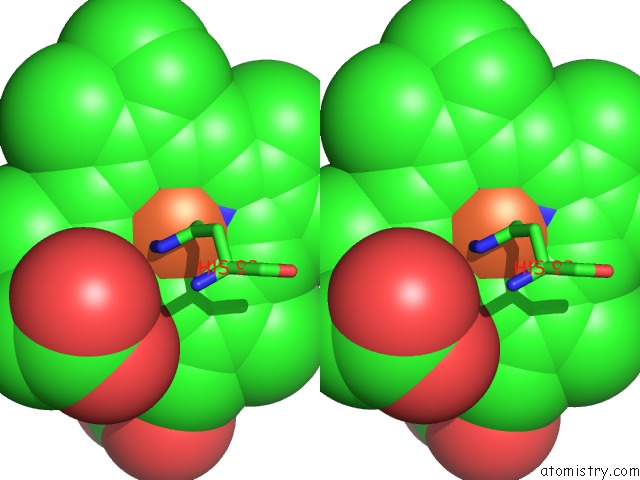
Stereo pair view
Mono view
Stereo pair view
A full contact list of Iron with other atoms in the Fe binding
site number 1 of Crystal Structure of Native Sperm Whale Myoglobin From Low Ionic Strength Enviroment (FORM2 ) within 5.0Å range:
|
Reference:
D.A.Kondrashov,
W.Zhang,
R.Aranda,
B.Stec,
G.N.Phillips Jr..
Sampling of the Native Conformational Ensemble of Myoglobin Via Structures in Different Crystalline Environments. Proteins V. 70 353 2008.
ISSN: ISSN 0887-3585
PubMed: 17680690
DOI: 10.1002/PROT.21499
Page generated: Sat Aug 3 15:25:02 2024
ISSN: ISSN 0887-3585
PubMed: 17680690
DOI: 10.1002/PROT.21499
Last articles
Cl in 2YNGCl in 2YNE
Cl in 2YNF
Cl in 2YNC
Cl in 2YLP
Cl in 2YN4
Cl in 2YM8
Cl in 2YKQ
Cl in 2YKO
Cl in 2YKP