Iron »
PDB 1tfz-1ubh »
1ub2 »
Iron in PDB 1ub2: Crystal Structure of Catalase-Peroxidase From Synechococcus Pcc 7942
Enzymatic activity of Crystal Structure of Catalase-Peroxidase From Synechococcus Pcc 7942
All present enzymatic activity of Crystal Structure of Catalase-Peroxidase From Synechococcus Pcc 7942:
1.11.1.6;
1.11.1.6;
Protein crystallography data
The structure of Crystal Structure of Catalase-Peroxidase From Synechococcus Pcc 7942, PDB code: 1ub2
was solved by
K.Wada,
T.Tada,
with X-Ray Crystallography technique. A brief refinement statistics is given in the table below:
| Resolution Low / High (Å) | 14.98 / 2.40 |
| Space group | P 41 21 2 |
| Cell size a, b, c (Å), α, β, γ (°) | 109.208, 109.208, 202.661, 90.00, 90.00, 90.00 |
| R / Rfree (%) | 19.9 / 23.2 |
Iron Binding Sites:
The binding sites of Iron atom in the Crystal Structure of Catalase-Peroxidase From Synechococcus Pcc 7942
(pdb code 1ub2). This binding sites where shown within
5.0 Angstroms radius around Iron atom.
In total only one binding site of Iron was determined in the Crystal Structure of Catalase-Peroxidase From Synechococcus Pcc 7942, PDB code: 1ub2:
In total only one binding site of Iron was determined in the Crystal Structure of Catalase-Peroxidase From Synechococcus Pcc 7942, PDB code: 1ub2:
Iron binding site 1 out of 1 in 1ub2
Go back to
Iron binding site 1 out
of 1 in the Crystal Structure of Catalase-Peroxidase From Synechococcus Pcc 7942
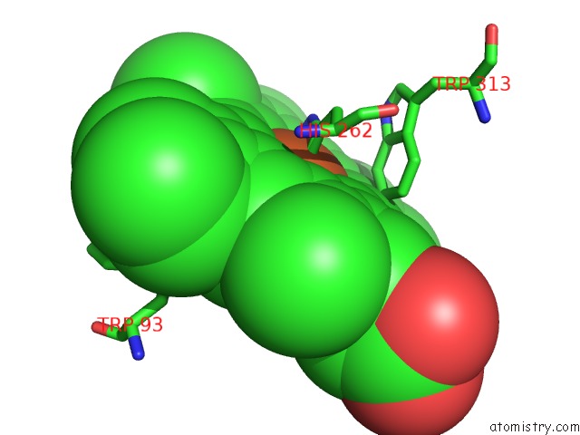
Mono view
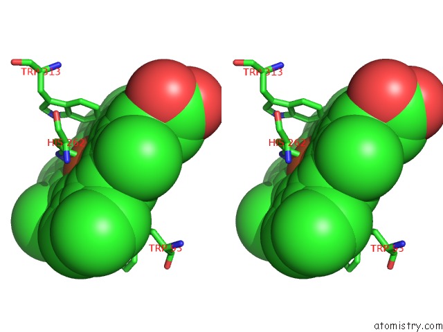
Stereo pair view
Mono view
Stereo pair view
A full contact list of Iron with other atoms in the Fe binding
site number 1 of Crystal Structure of Catalase-Peroxidase From Synechococcus Pcc 7942 within 5.0Å range:
|
Reference:
K.Wada,
T.Tada.
Crystal Structure of Catalase-Peroxidase From Synechococcus Pcc 7942 To Be Published.
Page generated: Sat Aug 3 15:26:08 2024
Last articles
F in 7NTHF in 7NTI
F in 7NPC
F in 7NRG
F in 7NR5
F in 7NQS
F in 7NOS
F in 7NP5
F in 7NDV
F in 7NP6