Iron »
PDB 1tfz-1ubh »
1ubb »
Iron in PDB 1ubb: Crystal Structure of Rat Ho-1 in Complex with Ferrous Heme
Enzymatic activity of Crystal Structure of Rat Ho-1 in Complex with Ferrous Heme
All present enzymatic activity of Crystal Structure of Rat Ho-1 in Complex with Ferrous Heme:
1.14.99.3;
1.14.99.3;
Protein crystallography data
The structure of Crystal Structure of Rat Ho-1 in Complex with Ferrous Heme, PDB code: 1ubb
was solved by
M.Sugishima,
H.Sakamoto,
Y.Higashimoto,
M.Noguchi,
K.Fukuyama,
with X-Ray Crystallography technique. A brief refinement statistics is given in the table below:
| Resolution Low / High (Å) | 40.20 / 2.30 |
| Space group | P 32 2 1 |
| Cell size a, b, c (Å), α, β, γ (°) | 65.300, 65.300, 120.500, 90.00, 90.00, 120.00 |
| R / Rfree (%) | 18.5 / 22 |
Iron Binding Sites:
The binding sites of Iron atom in the Crystal Structure of Rat Ho-1 in Complex with Ferrous Heme
(pdb code 1ubb). This binding sites where shown within
5.0 Angstroms radius around Iron atom.
In total only one binding site of Iron was determined in the Crystal Structure of Rat Ho-1 in Complex with Ferrous Heme, PDB code: 1ubb:
In total only one binding site of Iron was determined in the Crystal Structure of Rat Ho-1 in Complex with Ferrous Heme, PDB code: 1ubb:
Iron binding site 1 out of 1 in 1ubb
Go back to
Iron binding site 1 out
of 1 in the Crystal Structure of Rat Ho-1 in Complex with Ferrous Heme
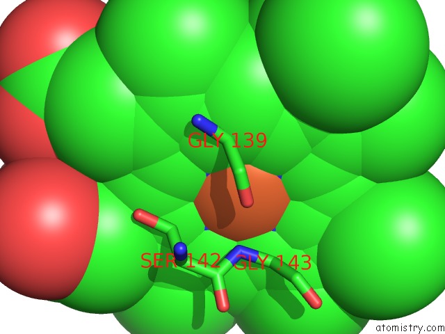
Mono view
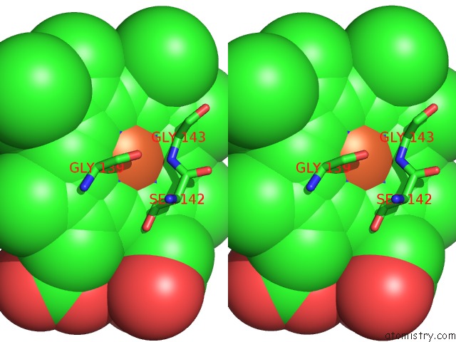
Stereo pair view
Mono view
Stereo pair view
A full contact list of Iron with other atoms in the Fe binding
site number 1 of Crystal Structure of Rat Ho-1 in Complex with Ferrous Heme within 5.0Å range:
|
Reference:
M.Sugishima,
H.Sakamoto,
M.Noguchi,
K.Fukuyama.
Crystal Structures of Ferrous and Co-, Cn(-)-, and No-Bound Forms of Rat Heme Oxygenase-1 (Ho-1) in Complex with Heme: Structural Implications For Discrimination Between Co and O(2) in Ho-1. Biochemistry V. 42 9898 2003.
ISSN: ISSN 0006-2960
PubMed: 12924938
DOI: 10.1021/BI027268I
Page generated: Sat Aug 3 15:26:09 2024
ISSN: ISSN 0006-2960
PubMed: 12924938
DOI: 10.1021/BI027268I
Last articles
Zn in 9J0NZn in 9J0O
Zn in 9J0P
Zn in 9FJX
Zn in 9EKB
Zn in 9C0F
Zn in 9CAH
Zn in 9CH0
Zn in 9CH3
Zn in 9CH1