Iron »
PDB 1ubj-1uvy »
1uob »
Iron in PDB 1uob: Deacetoxycephalosporin C Synthase Complexed with 2-Oxoglutarate and Penicillin G
Enzymatic activity of Deacetoxycephalosporin C Synthase Complexed with 2-Oxoglutarate and Penicillin G
All present enzymatic activity of Deacetoxycephalosporin C Synthase Complexed with 2-Oxoglutarate and Penicillin G:
1.14.20.1;
1.14.20.1;
Protein crystallography data
The structure of Deacetoxycephalosporin C Synthase Complexed with 2-Oxoglutarate and Penicillin G, PDB code: 1uob
was solved by
K.Valegard,
A.C.Terwisscha Van Scheltinga,
A.Dubus,
L.M.Oster,
G.Rhangino,
J.Hajdu,
I.Andersson,
with X-Ray Crystallography technique. A brief refinement statistics is given in the table below:
| Resolution Low / High (Å) | 56.80 / 1.70 |
| Space group | H 3 |
| Cell size a, b, c (Å), α, β, γ (°) | 106.600, 106.600, 71.600, 90.00, 90.00, 120.00 |
| R / Rfree (%) | 18.9 / 23.6 |
Iron Binding Sites:
The binding sites of Iron atom in the Deacetoxycephalosporin C Synthase Complexed with 2-Oxoglutarate and Penicillin G
(pdb code 1uob). This binding sites where shown within
5.0 Angstroms radius around Iron atom.
In total only one binding site of Iron was determined in the Deacetoxycephalosporin C Synthase Complexed with 2-Oxoglutarate and Penicillin G, PDB code: 1uob:
In total only one binding site of Iron was determined in the Deacetoxycephalosporin C Synthase Complexed with 2-Oxoglutarate and Penicillin G, PDB code: 1uob:
Iron binding site 1 out of 1 in 1uob
Go back to
Iron binding site 1 out
of 1 in the Deacetoxycephalosporin C Synthase Complexed with 2-Oxoglutarate and Penicillin G
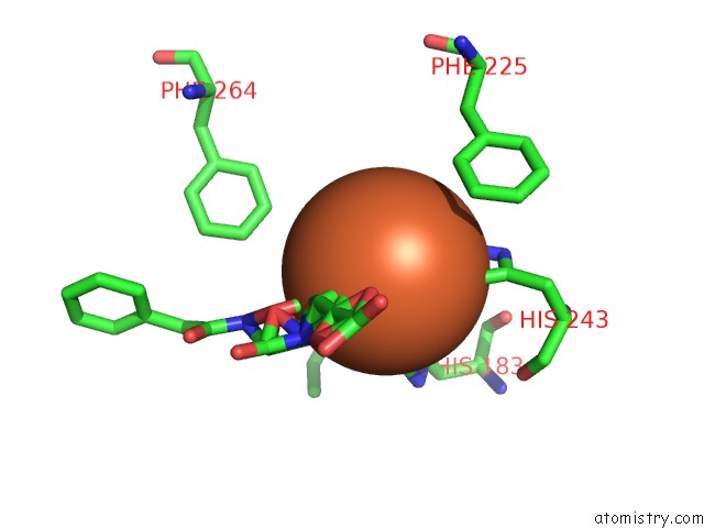
Mono view
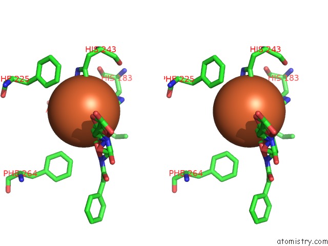
Stereo pair view
Mono view
Stereo pair view
A full contact list of Iron with other atoms in the Fe binding
site number 1 of Deacetoxycephalosporin C Synthase Complexed with 2-Oxoglutarate and Penicillin G within 5.0Å range:
|
Reference:
K.Valegard,
A.C.Terwisscha Van Scheltinga,
A.Dubus,
G.Ranghino,
L.M.Oster,
J.Hajdu,
I.Andersson.
The Structural Basis of Cephalosporin Formation in A Mononuclear Ferrous Enzyme Nat.Struct.Mol.Biol. V. 11 95 2004.
ISSN: ISSN 1545-9993
PubMed: 14718929
DOI: 10.1038/NSMB712
Page generated: Wed Jul 16 21:27:56 2025
ISSN: ISSN 1545-9993
PubMed: 14718929
DOI: 10.1038/NSMB712
Last articles
Fe in 2YXOFe in 2YRS
Fe in 2YXC
Fe in 2YNM
Fe in 2YVJ
Fe in 2YP1
Fe in 2YU2
Fe in 2YU1
Fe in 2YQB
Fe in 2YOO