Iron »
PDB 1ubj-1uvy »
1uog »
Iron in PDB 1uog: Deacetoxycephalosporin C Synthase Complexed with Deacetoxycephalosporin C
Enzymatic activity of Deacetoxycephalosporin C Synthase Complexed with Deacetoxycephalosporin C
All present enzymatic activity of Deacetoxycephalosporin C Synthase Complexed with Deacetoxycephalosporin C:
1.14.20.1;
1.14.20.1;
Protein crystallography data
The structure of Deacetoxycephalosporin C Synthase Complexed with Deacetoxycephalosporin C, PDB code: 1uog
was solved by
K.Valegard,
A.C.Terwisscha Van Scheltinga,
A.Dubus,
L.M.Oster,
G.Rhangino,
J.Hajdu,
I.Andersson,
with X-Ray Crystallography technique. A brief refinement statistics is given in the table below:
| Resolution Low / High (Å) | 25.40 / 1.70 |
| Space group | H 3 |
| Cell size a, b, c (Å), α, β, γ (°) | 106.600, 106.600, 74.000, 90.00, 90.00, 120.00 |
| R / Rfree (%) | 20.4 / 24.2 |
Iron Binding Sites:
The binding sites of Iron atom in the Deacetoxycephalosporin C Synthase Complexed with Deacetoxycephalosporin C
(pdb code 1uog). This binding sites where shown within
5.0 Angstroms radius around Iron atom.
In total only one binding site of Iron was determined in the Deacetoxycephalosporin C Synthase Complexed with Deacetoxycephalosporin C, PDB code: 1uog:
In total only one binding site of Iron was determined in the Deacetoxycephalosporin C Synthase Complexed with Deacetoxycephalosporin C, PDB code: 1uog:
Iron binding site 1 out of 1 in 1uog
Go back to
Iron binding site 1 out
of 1 in the Deacetoxycephalosporin C Synthase Complexed with Deacetoxycephalosporin C
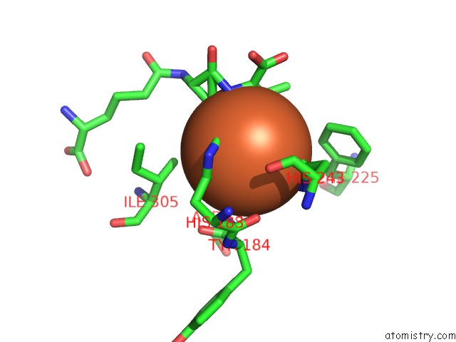
Mono view
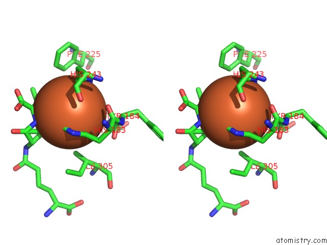
Stereo pair view
Mono view
Stereo pair view
A full contact list of Iron with other atoms in the Fe binding
site number 1 of Deacetoxycephalosporin C Synthase Complexed with Deacetoxycephalosporin C within 5.0Å range:
|
Reference:
K.Valegard,
A.C.Terwisscha Van Scheltinga,
A.Dubus,
G.Ranghino,
L.M.Oster,
J.Hajdu,
I.Andersson.
The Structural Basis of Cephalosporin Formation in A Mononuclear Ferrous Enzyme Nat.Struct.Mol.Biol. V. 11 95 2004.
ISSN: ISSN 1545-9993
PubMed: 14718929
DOI: 10.1038/NSMB712
Page generated: Sat Aug 3 15:55:05 2024
ISSN: ISSN 1545-9993
PubMed: 14718929
DOI: 10.1038/NSMB712
Last articles
Zn in 9MJ5Zn in 9HNW
Zn in 9G0L
Zn in 9FNE
Zn in 9DZN
Zn in 9E0I
Zn in 9D32
Zn in 9DAK
Zn in 8ZXC
Zn in 8ZUF