Iron »
PDB 1ubj-1uvy »
1uvh »
Iron in PDB 1uvh: X-Ray Structure of Dps From Mycobacterium Smegmatis
Protein crystallography data
The structure of X-Ray Structure of Dps From Mycobacterium Smegmatis, PDB code: 1uvh
was solved by
A.Ilari,
P.Ceci,
E.Falvo,
E.Chiancone,
with X-Ray Crystallography technique. A brief refinement statistics is given in the table below:
| Resolution Low / High (Å) | 40 / 2.8 |
| Space group | H 3 2 |
| Cell size a, b, c (Å), α, β, γ (°) | 124.300, 124.300, 304.650, 90.00, 90.00, 120.00 |
| R / Rfree (%) | 26 / 34 |
Iron Binding Sites:
The binding sites of Iron atom in the X-Ray Structure of Dps From Mycobacterium Smegmatis
(pdb code 1uvh). This binding sites where shown within
5.0 Angstroms radius around Iron atom.
In total 4 binding sites of Iron where determined in the X-Ray Structure of Dps From Mycobacterium Smegmatis, PDB code: 1uvh:
Jump to Iron binding site number: 1; 2; 3; 4;
In total 4 binding sites of Iron where determined in the X-Ray Structure of Dps From Mycobacterium Smegmatis, PDB code: 1uvh:
Jump to Iron binding site number: 1; 2; 3; 4;
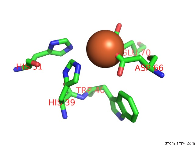
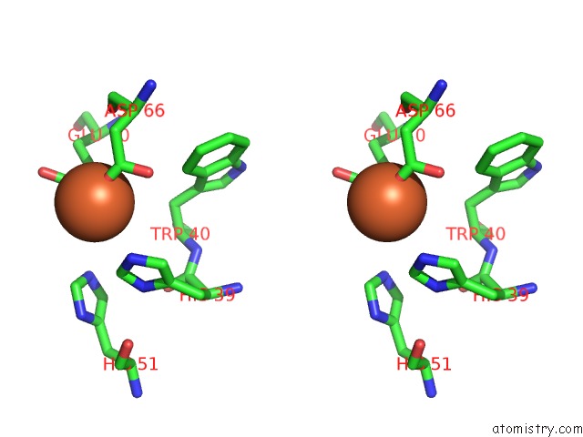
Iron binding site 1 out of 4 in 1uvh
Go back to
Iron binding site 1 out
of 4 in the X-Ray Structure of Dps From Mycobacterium Smegmatis
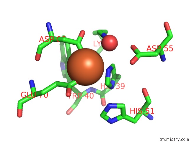
Mono view
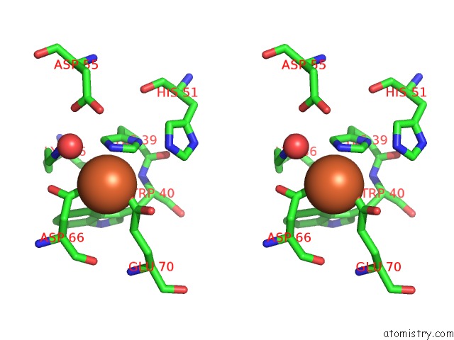
Stereo pair view
Mono view
Stereo pair view
A full contact list of Iron with other atoms in the Fe binding
site number 1 of X-Ray Structure of Dps From Mycobacterium Smegmatis within 5.0Å range:
|
Iron binding site 2 out of 4 in 1uvh
Go back to
Iron binding site 2 out
of 4 in the X-Ray Structure of Dps From Mycobacterium Smegmatis
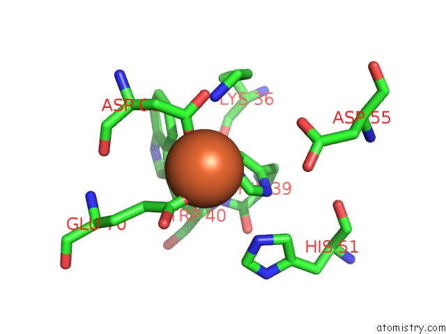
Mono view
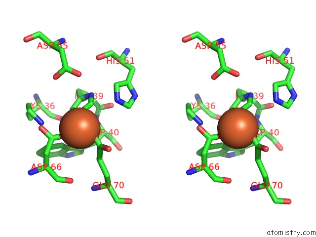
Stereo pair view
Mono view
Stereo pair view
A full contact list of Iron with other atoms in the Fe binding
site number 2 of X-Ray Structure of Dps From Mycobacterium Smegmatis within 5.0Å range:
|
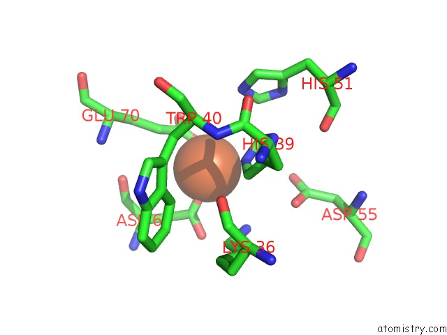
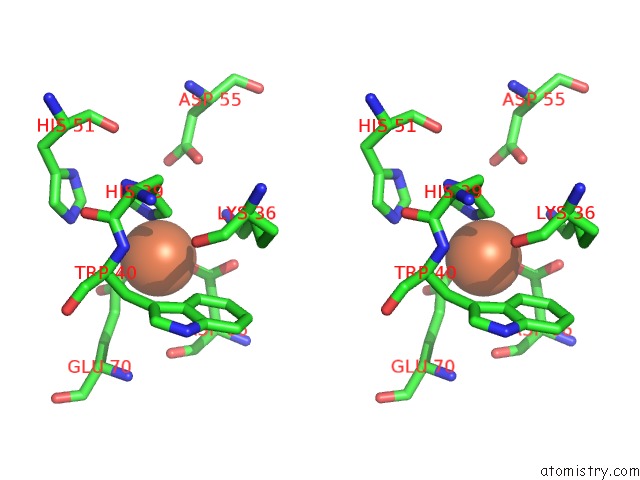
Iron binding site 3 out of 4 in 1uvh
Go back to
Iron binding site 3 out
of 4 in the X-Ray Structure of Dps From Mycobacterium Smegmatis
Mono view
Stereo pair view
Mono view
Stereo pair view
A full contact list of Iron with other atoms in the Fe binding
site number 3 of X-Ray Structure of Dps From Mycobacterium Smegmatis within 5.0Å range:
|
Iron binding site 4 out of 4 in 1uvh
Go back to
Iron binding site 4 out
of 4 in the X-Ray Structure of Dps From Mycobacterium Smegmatis
Mono view
Stereo pair view
Mono view
Stereo pair view
A full contact list of Iron with other atoms in the Fe binding
site number 4 of X-Ray Structure of Dps From Mycobacterium Smegmatis within 5.0Å range:
|
Reference:
P.Ceci,
A.Ilari,
E.Falvo,
L.Giangiacomo,
E.Chiancone.
Reassessment of Protein Stability, Dna Binding, and Protection of Mycobacterium Smegmatis Dps. J.Biol.Chem. V. 280 34776 2005.
ISSN: ISSN 0021-9258
PubMed: 16030020
DOI: 10.1074/JBC.M502343200
Page generated: Wed Jul 16 21:30:51 2025
ISSN: ISSN 0021-9258
PubMed: 16030020
DOI: 10.1074/JBC.M502343200
Last articles
Fe in 2YXOFe in 2YRS
Fe in 2YXC
Fe in 2YNM
Fe in 2YVJ
Fe in 2YP1
Fe in 2YU2
Fe in 2YU1
Fe in 2YQB
Fe in 2YOO