Iron »
PDB 1uwm-1vme »
1uwm »
Iron in PDB 1uwm: Reduced Ferredoxin 6 From Rhodobacter Capsulatus
Protein crystallography data
The structure of Reduced Ferredoxin 6 From Rhodobacter Capsulatus, PDB code: 1uwm
was solved by
G.Sainz,
J.Jakoncic,
L.C.Sieker,
V.Stojanoff,
N.Sanishvili,
M.Asso,
P.Bertrand,
J.Armengaud,
Y.Jouanneau,
with X-Ray Crystallography technique. A brief refinement statistics is given in the table below:
| Resolution Low / High (Å) | 28.83 / 2.00 |
| Space group | P 21 21 21 |
| Cell size a, b, c (Å), α, β, γ (°) | 45.542, 50.335, 55.349, 90.00, 90.00, 90.00 |
| R / Rfree (%) | 23 / 25.7 |
Iron Binding Sites:
The binding sites of Iron atom in the Reduced Ferredoxin 6 From Rhodobacter Capsulatus
(pdb code 1uwm). This binding sites where shown within
5.0 Angstroms radius around Iron atom.
In total 2 binding sites of Iron where determined in the Reduced Ferredoxin 6 From Rhodobacter Capsulatus, PDB code: 1uwm:
Jump to Iron binding site number: 1; 2;
In total 2 binding sites of Iron where determined in the Reduced Ferredoxin 6 From Rhodobacter Capsulatus, PDB code: 1uwm:
Jump to Iron binding site number: 1; 2;
Iron binding site 1 out of 2 in 1uwm
Go back to
Iron binding site 1 out
of 2 in the Reduced Ferredoxin 6 From Rhodobacter Capsulatus
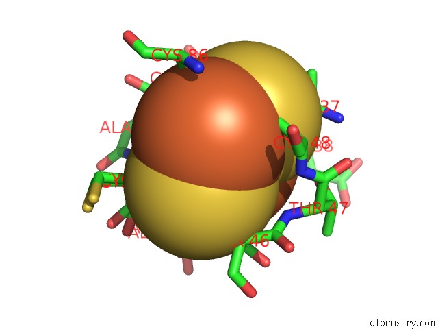
Mono view
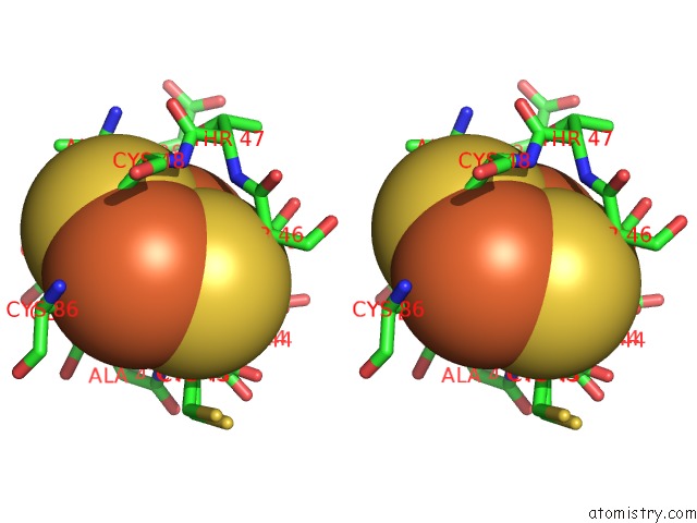
Stereo pair view
Mono view
Stereo pair view
A full contact list of Iron with other atoms in the Fe binding
site number 1 of Reduced Ferredoxin 6 From Rhodobacter Capsulatus within 5.0Å range:
|
Iron binding site 2 out of 2 in 1uwm
Go back to
Iron binding site 2 out
of 2 in the Reduced Ferredoxin 6 From Rhodobacter Capsulatus
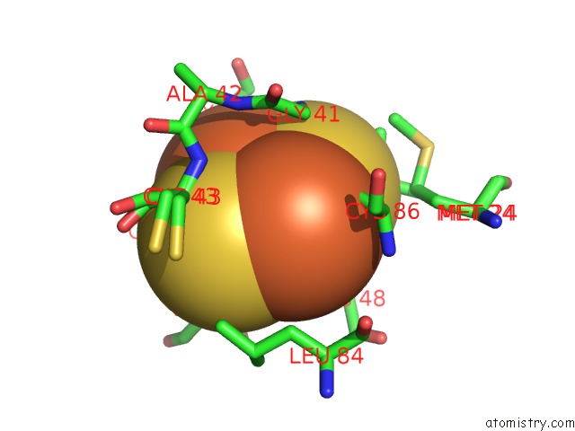
Mono view
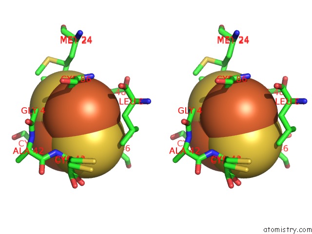
Stereo pair view
Mono view
Stereo pair view
A full contact list of Iron with other atoms in the Fe binding
site number 2 of Reduced Ferredoxin 6 From Rhodobacter Capsulatus within 5.0Å range:
|
Reference:
G.Sainz,
J.Jakoncic,
L.C.Sieker,
V.Stojanoff,
N.Sanishvili,
M.Asso,
P.Bertrand,
J.Armengaud,
Y.Jouanneau.
Structure of A [2FE-2S] Ferredoxin From Rhodobacter Capsulatus Likely Involved in Fe-S Cluster Biogenesis and Conformational Changes Observed Upon Reduction. J.Biol.Inorg.Chem. V. 11 235 2006.
ISSN: ISSN 0949-8257
PubMed: 16402206
DOI: 10.1007/S00775-005-0069-2
Page generated: Sat Aug 3 16:07:00 2024
ISSN: ISSN 0949-8257
PubMed: 16402206
DOI: 10.1007/S00775-005-0069-2
Last articles
Zn in 9MJ5Zn in 9HNW
Zn in 9G0L
Zn in 9FNE
Zn in 9DZN
Zn in 9E0I
Zn in 9D32
Zn in 9DAK
Zn in 8ZXC
Zn in 8ZUF