Iron »
PDB 1uwm-1vme »
1uzr »
Iron in PDB 1uzr: Crystal Structure of the Class Ib Ribonucleotide Reductase R2F-2 Subunit From Mycobacterium Tuberculosis
Enzymatic activity of Crystal Structure of the Class Ib Ribonucleotide Reductase R2F-2 Subunit From Mycobacterium Tuberculosis
All present enzymatic activity of Crystal Structure of the Class Ib Ribonucleotide Reductase R2F-2 Subunit From Mycobacterium Tuberculosis:
1.17.4.1;
1.17.4.1;
Protein crystallography data
The structure of Crystal Structure of the Class Ib Ribonucleotide Reductase R2F-2 Subunit From Mycobacterium Tuberculosis, PDB code: 1uzr
was solved by
M.Uppsten,
J.Davis,
H.Rubin,
U.Uhlin,
with X-Ray Crystallography technique. A brief refinement statistics is given in the table below:
| Resolution Low / High (Å) | 111.80 / 2.2 |
| Space group | P 42 21 2 |
| Cell size a, b, c (Å), α, β, γ (°) | 161.492, 161.492, 115.530, 90.00, 90.00, 90.00 |
| R / Rfree (%) | 17.5 / 20.2 |
Iron Binding Sites:
The binding sites of Iron atom in the Crystal Structure of the Class Ib Ribonucleotide Reductase R2F-2 Subunit From Mycobacterium Tuberculosis
(pdb code 1uzr). This binding sites where shown within
5.0 Angstroms radius around Iron atom.
In total 6 binding sites of Iron where determined in the Crystal Structure of the Class Ib Ribonucleotide Reductase R2F-2 Subunit From Mycobacterium Tuberculosis, PDB code: 1uzr:
Jump to Iron binding site number: 1; 2; 3; 4; 5; 6;
In total 6 binding sites of Iron where determined in the Crystal Structure of the Class Ib Ribonucleotide Reductase R2F-2 Subunit From Mycobacterium Tuberculosis, PDB code: 1uzr:
Jump to Iron binding site number: 1; 2; 3; 4; 5; 6;
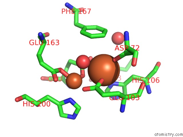
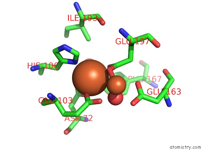
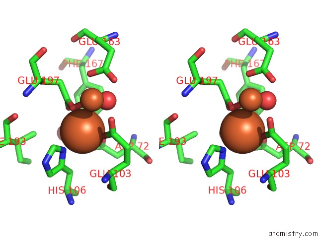
Iron binding site 1 out of 6 in 1uzr
Go back to
Iron binding site 1 out
of 6 in the Crystal Structure of the Class Ib Ribonucleotide Reductase R2F-2 Subunit From Mycobacterium Tuberculosis
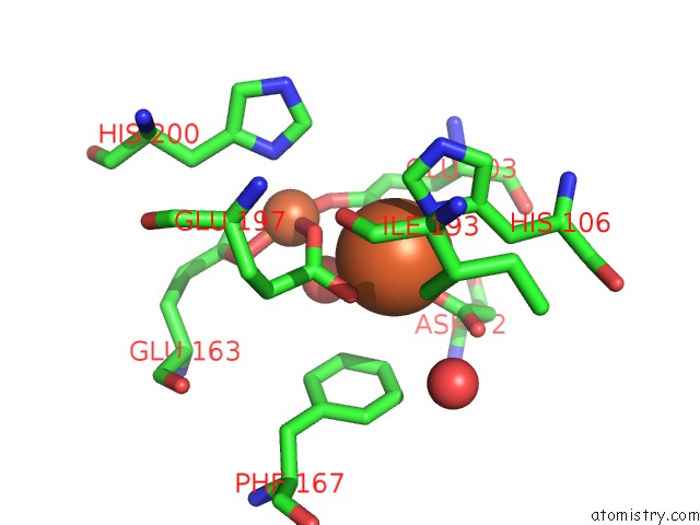
Mono view
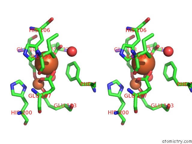
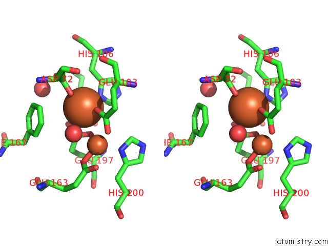
Stereo pair view
Mono view
Stereo pair view
A full contact list of Iron with other atoms in the Fe binding
site number 1 of Crystal Structure of the Class Ib Ribonucleotide Reductase R2F-2 Subunit From Mycobacterium Tuberculosis within 5.0Å range:
|
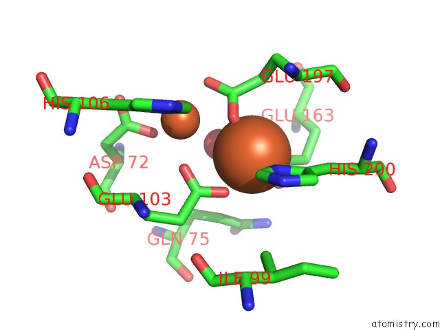
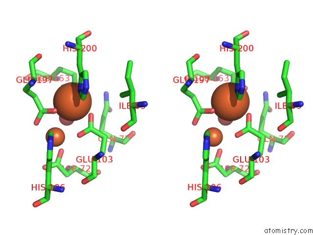
Iron binding site 2 out of 6 in 1uzr
Go back to
Iron binding site 2 out
of 6 in the Crystal Structure of the Class Ib Ribonucleotide Reductase R2F-2 Subunit From Mycobacterium Tuberculosis
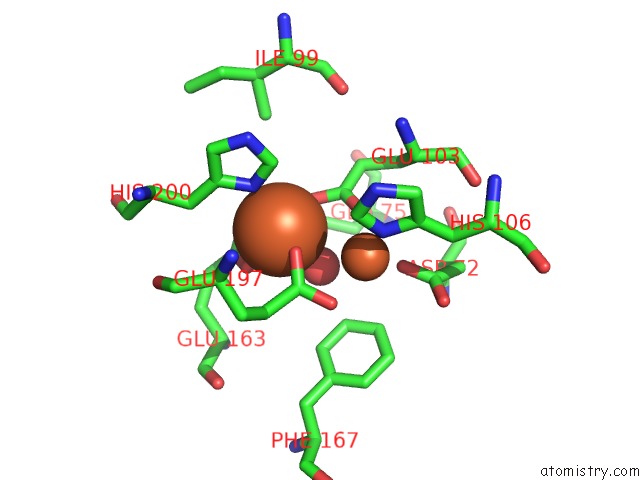
Mono view
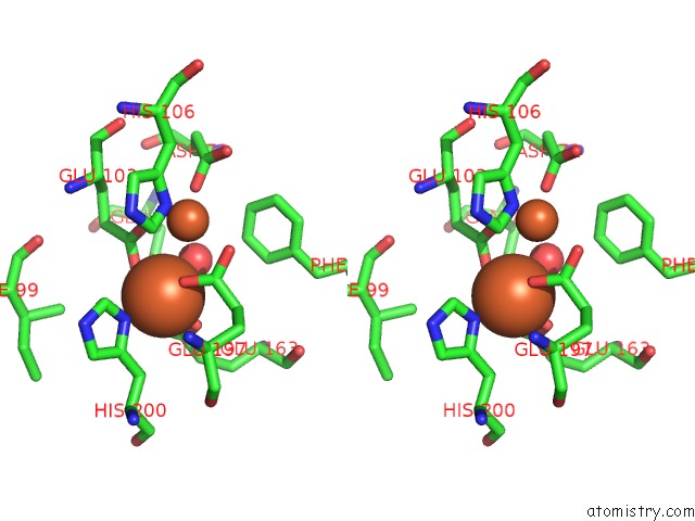
Stereo pair view
Mono view
Stereo pair view
A full contact list of Iron with other atoms in the Fe binding
site number 2 of Crystal Structure of the Class Ib Ribonucleotide Reductase R2F-2 Subunit From Mycobacterium Tuberculosis within 5.0Å range:
|
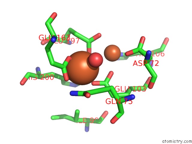
Iron binding site 3 out of 6 in 1uzr
Go back to
Iron binding site 3 out
of 6 in the Crystal Structure of the Class Ib Ribonucleotide Reductase R2F-2 Subunit From Mycobacterium Tuberculosis
Mono view
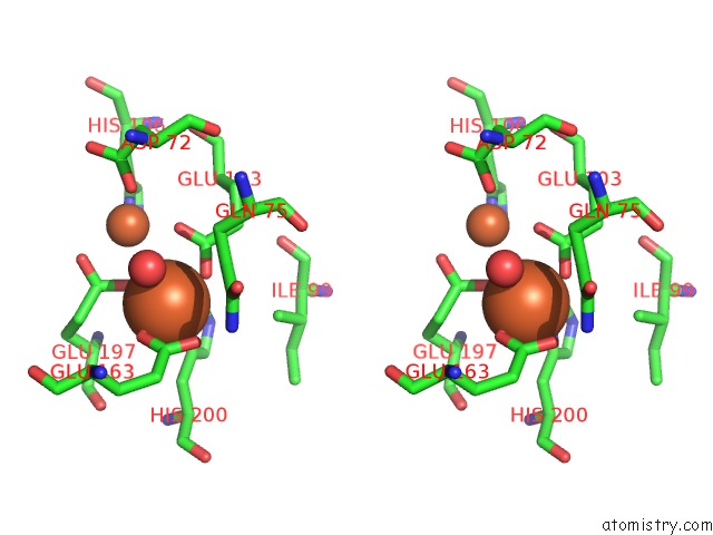
Stereo pair view
Mono view
Stereo pair view
A full contact list of Iron with other atoms in the Fe binding
site number 3 of Crystal Structure of the Class Ib Ribonucleotide Reductase R2F-2 Subunit From Mycobacterium Tuberculosis within 5.0Å range:
|
Iron binding site 4 out of 6 in 1uzr
Go back to
Iron binding site 4 out
of 6 in the Crystal Structure of the Class Ib Ribonucleotide Reductase R2F-2 Subunit From Mycobacterium Tuberculosis
Mono view
Stereo pair view
Mono view
Stereo pair view
A full contact list of Iron with other atoms in the Fe binding
site number 4 of Crystal Structure of the Class Ib Ribonucleotide Reductase R2F-2 Subunit From Mycobacterium Tuberculosis within 5.0Å range:
|
Iron binding site 5 out of 6 in 1uzr
Go back to
Iron binding site 5 out
of 6 in the Crystal Structure of the Class Ib Ribonucleotide Reductase R2F-2 Subunit From Mycobacterium Tuberculosis
Mono view
Stereo pair view
Mono view
Stereo pair view
A full contact list of Iron with other atoms in the Fe binding
site number 5 of Crystal Structure of the Class Ib Ribonucleotide Reductase R2F-2 Subunit From Mycobacterium Tuberculosis within 5.0Å range:
|
Iron binding site 6 out of 6 in 1uzr
Go back to
Iron binding site 6 out
of 6 in the Crystal Structure of the Class Ib Ribonucleotide Reductase R2F-2 Subunit From Mycobacterium Tuberculosis
Mono view
Stereo pair view
Mono view
Stereo pair view
A full contact list of Iron with other atoms in the Fe binding
site number 6 of Crystal Structure of the Class Ib Ribonucleotide Reductase R2F-2 Subunit From Mycobacterium Tuberculosis within 5.0Å range:
|
Reference:
M.Uppsten,
J.Davis,
H.Rubin,
U.Uhlin.
Crystal Structure of the Biologically Active Form of Class Ib Ribonucleotide Reductase Small Subunit From Mycobacterium Tuberculosis Febs Lett. V. 569 117 2004.
ISSN: ISSN 0014-5793
PubMed: 15225619
DOI: 10.1016/J.FEBSLET.2004.05.059
Page generated: Wed Jul 16 21:34:24 2025
ISSN: ISSN 0014-5793
PubMed: 15225619
DOI: 10.1016/J.FEBSLET.2004.05.059
Last articles
Fe in 2YXOFe in 2YRS
Fe in 2YXC
Fe in 2YNM
Fe in 2YVJ
Fe in 2YP1
Fe in 2YU2
Fe in 2YU1
Fe in 2YQB
Fe in 2YOO