Iron »
PDB 1uwm-1vme »
1v0h »
Iron in PDB 1v0h: Ascobate Peroxidase From Soybean Cytosol in Complex with Salicylhydroxamic Acid
Enzymatic activity of Ascobate Peroxidase From Soybean Cytosol in Complex with Salicylhydroxamic Acid
All present enzymatic activity of Ascobate Peroxidase From Soybean Cytosol in Complex with Salicylhydroxamic Acid:
1.11.1.11;
1.11.1.11;
Protein crystallography data
The structure of Ascobate Peroxidase From Soybean Cytosol in Complex with Salicylhydroxamic Acid, PDB code: 1v0h
was solved by
K.H.Sharp,
E.L.Raven,
P.C.E.Moody,
with X-Ray Crystallography technique. A brief refinement statistics is given in the table below:
| Resolution Low / High (Å) | 55.90 / 1.46 |
| Space group | P 42 21 2 |
| Cell size a, b, c (Å), α, β, γ (°) | 82.770, 82.770, 74.991, 90.00, 90.00, 90.00 |
| R / Rfree (%) | 15 / 18.2 |
Other elements in 1v0h:
The structure of Ascobate Peroxidase From Soybean Cytosol in Complex with Salicylhydroxamic Acid also contains other interesting chemical elements:
| Sodium | (Na) | 1 atom |
Iron Binding Sites:
The binding sites of Iron atom in the Ascobate Peroxidase From Soybean Cytosol in Complex with Salicylhydroxamic Acid
(pdb code 1v0h). This binding sites where shown within
5.0 Angstroms radius around Iron atom.
In total only one binding site of Iron was determined in the Ascobate Peroxidase From Soybean Cytosol in Complex with Salicylhydroxamic Acid, PDB code: 1v0h:
In total only one binding site of Iron was determined in the Ascobate Peroxidase From Soybean Cytosol in Complex with Salicylhydroxamic Acid, PDB code: 1v0h:
Iron binding site 1 out of 1 in 1v0h
Go back to
Iron binding site 1 out
of 1 in the Ascobate Peroxidase From Soybean Cytosol in Complex with Salicylhydroxamic Acid
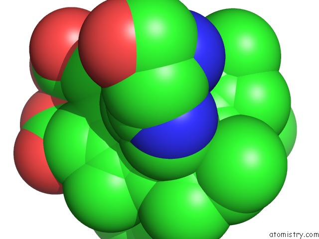
Mono view
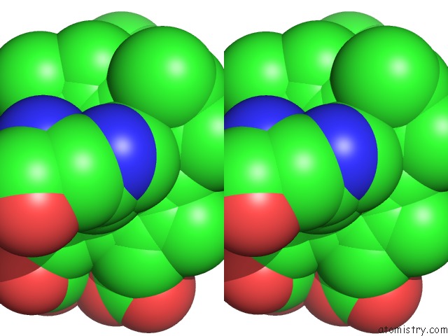
Stereo pair view
Mono view
Stereo pair view
A full contact list of Iron with other atoms in the Fe binding
site number 1 of Ascobate Peroxidase From Soybean Cytosol in Complex with Salicylhydroxamic Acid within 5.0Å range:
|
Reference:
K.H.Sharp,
P.C.E.Moody,
K.A.Brown,
E.L.Raven.
Crystal Structure of the Ascorbate Peroxidase-Salicylhydroxamic Acid Complex Biochemistry V. 43 8644 2004.
ISSN: ISSN 0006-2960
PubMed: 15236572
DOI: 10.1021/BI049343Q
Page generated: Sat Aug 3 16:07:00 2024
ISSN: ISSN 0006-2960
PubMed: 15236572
DOI: 10.1021/BI049343Q
Last articles
Zn in 9J0NZn in 9J0O
Zn in 9J0P
Zn in 9FJX
Zn in 9EKB
Zn in 9C0F
Zn in 9CAH
Zn in 9CH0
Zn in 9CH3
Zn in 9CH1