Iron »
PDB 1uwm-1vme »
1vck »
Iron in PDB 1vck: Crystal Structure of Ferredoxin Component of Carbazole 1,9A- Dioxygenase of Pseudomonas Resinovorans Strain CA10
Protein crystallography data
The structure of Crystal Structure of Ferredoxin Component of Carbazole 1,9A- Dioxygenase of Pseudomonas Resinovorans Strain CA10, PDB code: 1vck
was solved by
J.-W.Nam,
H.Noguchi,
Z.Fujiomoto,
H.Mizuno,
S.Fushinobu,
N.Kobashi,
K.Iwata,
T.Yoshida,
H.Habe,
H.Yamane,
T.Omori,
H.Nojiri,
with X-Ray Crystallography technique. A brief refinement statistics is given in the table below:
| Resolution Low / High (Å) | 29.63 / 1.90 |
| Space group | P 41 3 2 |
| Cell size a, b, c (Å), α, β, γ (°) | 98.281, 98.281, 98.281, 90.00, 90.00, 90.00 |
| R / Rfree (%) | 20.2 / 23.6 |
Iron Binding Sites:
The binding sites of Iron atom in the Crystal Structure of Ferredoxin Component of Carbazole 1,9A- Dioxygenase of Pseudomonas Resinovorans Strain CA10
(pdb code 1vck). This binding sites where shown within
5.0 Angstroms radius around Iron atom.
In total 3 binding sites of Iron where determined in the Crystal Structure of Ferredoxin Component of Carbazole 1,9A- Dioxygenase of Pseudomonas Resinovorans Strain CA10, PDB code: 1vck:
Jump to Iron binding site number: 1; 2; 3;
In total 3 binding sites of Iron where determined in the Crystal Structure of Ferredoxin Component of Carbazole 1,9A- Dioxygenase of Pseudomonas Resinovorans Strain CA10, PDB code: 1vck:
Jump to Iron binding site number: 1; 2; 3;
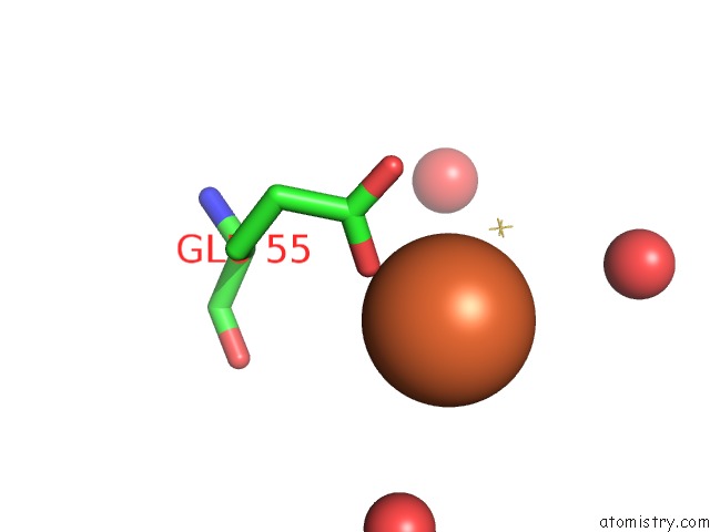
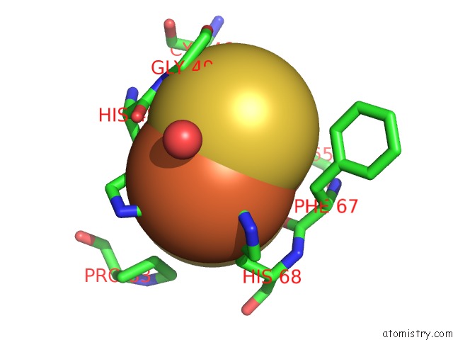
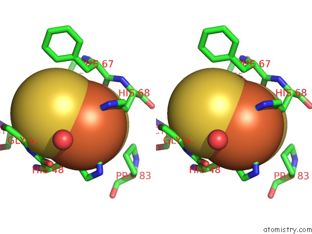
Iron binding site 1 out of 3 in 1vck
Go back to
Iron binding site 1 out
of 3 in the Crystal Structure of Ferredoxin Component of Carbazole 1,9A- Dioxygenase of Pseudomonas Resinovorans Strain CA10
Mono view
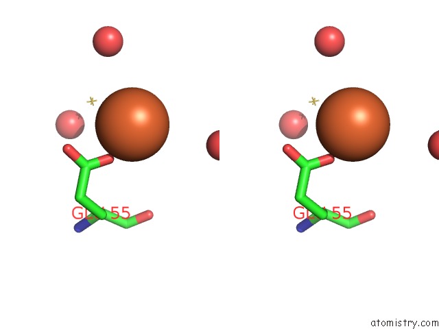
Stereo pair view
Mono view
Stereo pair view
A full contact list of Iron with other atoms in the Fe binding
site number 1 of Crystal Structure of Ferredoxin Component of Carbazole 1,9A- Dioxygenase of Pseudomonas Resinovorans Strain CA10 within 5.0Å range:
|
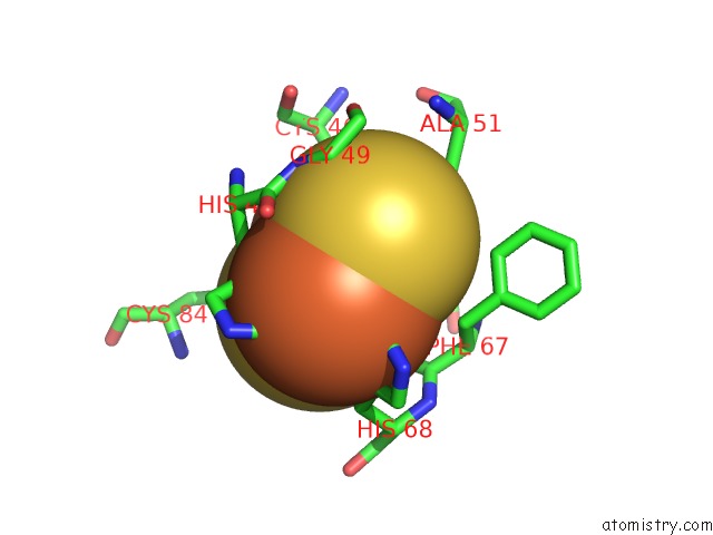
Iron binding site 2 out of 3 in 1vck
Go back to
Iron binding site 2 out
of 3 in the Crystal Structure of Ferredoxin Component of Carbazole 1,9A- Dioxygenase of Pseudomonas Resinovorans Strain CA10
Mono view
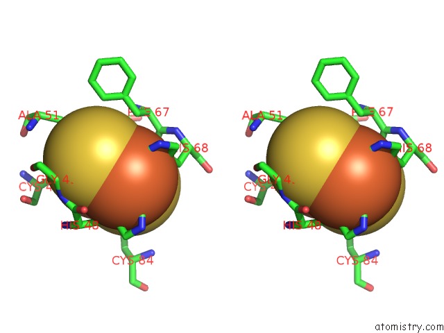
Stereo pair view
Mono view
Stereo pair view
A full contact list of Iron with other atoms in the Fe binding
site number 2 of Crystal Structure of Ferredoxin Component of Carbazole 1,9A- Dioxygenase of Pseudomonas Resinovorans Strain CA10 within 5.0Å range:
|
Iron binding site 3 out of 3 in 1vck
Go back to
Iron binding site 3 out
of 3 in the Crystal Structure of Ferredoxin Component of Carbazole 1,9A- Dioxygenase of Pseudomonas Resinovorans Strain CA10
Mono view
Stereo pair view
Mono view
Stereo pair view
A full contact list of Iron with other atoms in the Fe binding
site number 3 of Crystal Structure of Ferredoxin Component of Carbazole 1,9A- Dioxygenase of Pseudomonas Resinovorans Strain CA10 within 5.0Å range:
|
Reference:
J.-W.Nam,
H.Noguchi,
Z.Fujiomoto,
H.Mizuno,
Y.Ashikawa,
M.Abo,
S.Fushinobu,
N.Kobashi,
T.Wakagi,
K.Iwata,
T.Yoshida,
H.Habe,
H.Yamane,
T.Omori,
H.Nojiri.
Crystal Structure of the Ferredoxin Component of Carbazole 1,9A-Dioxygenase of Pseudomonas Resinovorans Strain CA10, A Novel Rieske Non-Heme Iron Oxygenase System Proteins V. 58 779 2005.
ISSN: ISSN 0887-3585
PubMed: 15645447
DOI: 10.1002/PROT.20374
Page generated: Sat Aug 3 16:12:01 2024
ISSN: ISSN 0887-3585
PubMed: 15645447
DOI: 10.1002/PROT.20374
Last articles
Zn in 9J0NZn in 9J0O
Zn in 9J0P
Zn in 9FJX
Zn in 9EKB
Zn in 9C0F
Zn in 9CAH
Zn in 9CH0
Zn in 9CH3
Zn in 9CH1