Iron »
PDB 1uwm-1vme »
1vlb »
Iron in PDB 1vlb: Structure Refinement of the Aldehyde Oxidoreductase From Desulfovibrio Gigas at 1.28 A
Enzymatic activity of Structure Refinement of the Aldehyde Oxidoreductase From Desulfovibrio Gigas at 1.28 A
All present enzymatic activity of Structure Refinement of the Aldehyde Oxidoreductase From Desulfovibrio Gigas at 1.28 A:
1.2.3.1;
1.2.3.1;
Protein crystallography data
The structure of Structure Refinement of the Aldehyde Oxidoreductase From Desulfovibrio Gigas at 1.28 A, PDB code: 1vlb
was solved by
J.M.Rebelo,
J.M.Dias,
R.Huber,
J.J.G.Moura,
M.J.Romao,
with X-Ray Crystallography technique. A brief refinement statistics is given in the table below:
| Resolution Low / High (Å) | 24.40 / 1.28 |
| Space group | P 61 2 2 |
| Cell size a, b, c (Å), α, β, γ (°) | 141.780, 141.780, 160.870, 90.00, 90.00, 120.00 |
| R / Rfree (%) | 14.8 / 19.3 |
Other elements in 1vlb:
The structure of Structure Refinement of the Aldehyde Oxidoreductase From Desulfovibrio Gigas at 1.28 A also contains other interesting chemical elements:
| Molybdenum | (Mo) | 1 atom |
| Magnesium | (Mg) | 2 atoms |
| Chlorine | (Cl) | 3 atoms |
Iron Binding Sites:
The binding sites of Iron atom in the Structure Refinement of the Aldehyde Oxidoreductase From Desulfovibrio Gigas at 1.28 A
(pdb code 1vlb). This binding sites where shown within
5.0 Angstroms radius around Iron atom.
In total 4 binding sites of Iron where determined in the Structure Refinement of the Aldehyde Oxidoreductase From Desulfovibrio Gigas at 1.28 A, PDB code: 1vlb:
Jump to Iron binding site number: 1; 2; 3; 4;
In total 4 binding sites of Iron where determined in the Structure Refinement of the Aldehyde Oxidoreductase From Desulfovibrio Gigas at 1.28 A, PDB code: 1vlb:
Jump to Iron binding site number: 1; 2; 3; 4;
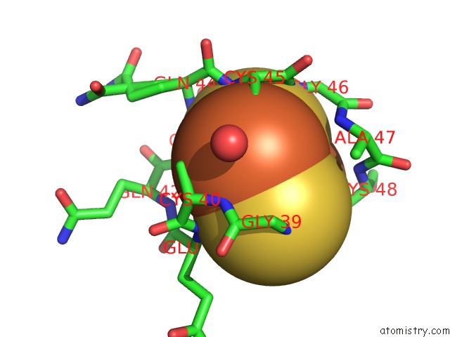
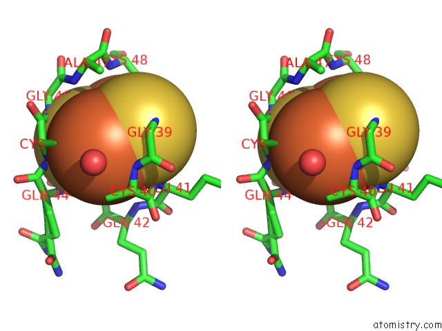
Iron binding site 1 out of 4 in 1vlb
Go back to
Iron binding site 1 out
of 4 in the Structure Refinement of the Aldehyde Oxidoreductase From Desulfovibrio Gigas at 1.28 A
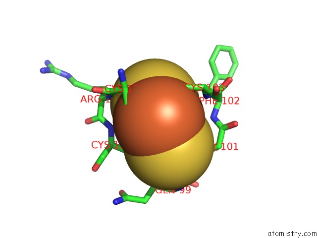
Mono view
Stereo pair view
Mono view
Stereo pair view
A full contact list of Iron with other atoms in the Fe binding
site number 1 of Structure Refinement of the Aldehyde Oxidoreductase From Desulfovibrio Gigas at 1.28 A within 5.0Å range:
|
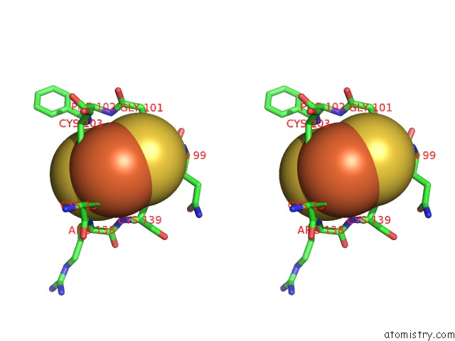
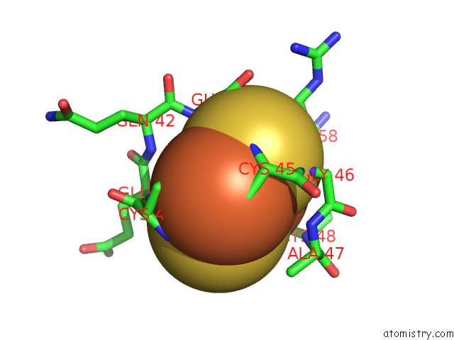
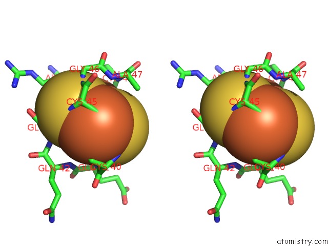
Iron binding site 2 out of 4 in 1vlb
Go back to
Iron binding site 2 out
of 4 in the Structure Refinement of the Aldehyde Oxidoreductase From Desulfovibrio Gigas at 1.28 A
Mono view
Stereo pair view
Mono view
Stereo pair view
A full contact list of Iron with other atoms in the Fe binding
site number 2 of Structure Refinement of the Aldehyde Oxidoreductase From Desulfovibrio Gigas at 1.28 A within 5.0Å range:
|
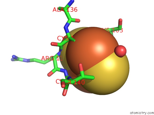
Iron binding site 3 out of 4 in 1vlb
Go back to
Iron binding site 3 out
of 4 in the Structure Refinement of the Aldehyde Oxidoreductase From Desulfovibrio Gigas at 1.28 A
Mono view
Stereo pair view
Mono view
Stereo pair view
A full contact list of Iron with other atoms in the Fe binding
site number 3 of Structure Refinement of the Aldehyde Oxidoreductase From Desulfovibrio Gigas at 1.28 A within 5.0Å range:
|
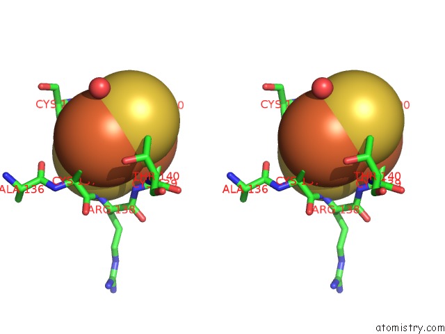
Iron binding site 4 out of 4 in 1vlb
Go back to
Iron binding site 4 out
of 4 in the Structure Refinement of the Aldehyde Oxidoreductase From Desulfovibrio Gigas at 1.28 A
Mono view
Stereo pair view
Mono view
Stereo pair view
A full contact list of Iron with other atoms in the Fe binding
site number 4 of Structure Refinement of the Aldehyde Oxidoreductase From Desulfovibrio Gigas at 1.28 A within 5.0Å range:
|
Reference:
J.M.Rebelo,
J.M.Dias,
R.Huber,
J.J.G.Moura,
M.J.Romao.
Structure Refinement of the Aldehyde Oxidoreductase From Desulfovibrio Gigas (Mop) at 1.28 A J.Biol.Inorg.Chem. V. 6 791 2001.
ISSN: ISSN 0949-8257
PubMed: 11713686
DOI: 10.1007/S007750100255
Page generated: Sat Aug 3 16:14:38 2024
ISSN: ISSN 0949-8257
PubMed: 11713686
DOI: 10.1007/S007750100255
Last articles
Cl in 5XIHCl in 5XHO
Cl in 5XIF
Cl in 5XHN
Cl in 5XHM
Cl in 5XGP
Cl in 5XH7
Cl in 5XH6
Cl in 5XGO
Cl in 5XD5