Iron »
PDB 1vrb-1w9m »
1w2l »
Iron in PDB 1w2l: Cytochrome C Domain of CAA3 Oxygen Oxidoreductase
Enzymatic activity of Cytochrome C Domain of CAA3 Oxygen Oxidoreductase
All present enzymatic activity of Cytochrome C Domain of CAA3 Oxygen Oxidoreductase:
1.9.3.1;
1.9.3.1;
Protein crystallography data
The structure of Cytochrome C Domain of CAA3 Oxygen Oxidoreductase, PDB code: 1w2l
was solved by
V.Srinivasan,
C.Rajendran,
F.L.Sousa,
A.M.P.Melo,
L.M.Saraiva,
M.M.Pereira,
M.Santana,
M.Teixeira,
H.Michel,
with X-Ray Crystallography technique. A brief refinement statistics is given in the table below:
| Resolution Low / High (Å) | 10.00 / 1.30 |
| Space group | P 41 21 2 |
| Cell size a, b, c (Å), α, β, γ (°) | 54.049, 54.049, 91.347, 90.00, 90.00, 90.00 |
| R / Rfree (%) | 14.2 / 17 |
Iron Binding Sites:
The binding sites of Iron atom in the Cytochrome C Domain of CAA3 Oxygen Oxidoreductase
(pdb code 1w2l). This binding sites where shown within
5.0 Angstroms radius around Iron atom.
In total only one binding site of Iron was determined in the Cytochrome C Domain of CAA3 Oxygen Oxidoreductase, PDB code: 1w2l:
In total only one binding site of Iron was determined in the Cytochrome C Domain of CAA3 Oxygen Oxidoreductase, PDB code: 1w2l:
Iron binding site 1 out of 1 in 1w2l
Go back to
Iron binding site 1 out
of 1 in the Cytochrome C Domain of CAA3 Oxygen Oxidoreductase
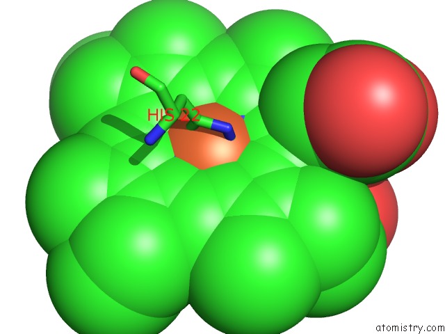
Mono view
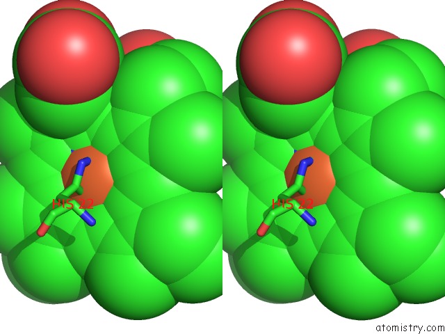
Stereo pair view
Mono view
Stereo pair view
A full contact list of Iron with other atoms in the Fe binding
site number 1 of Cytochrome C Domain of CAA3 Oxygen Oxidoreductase within 5.0Å range:
|
Reference:
V.Srinivasan,
C.Rajendran,
F.L.Sousa,
A.M.Melo,
L.M.Saraiva,
M.M.Pereira,
M.Santana,
M.Teixeira,
H.Michel.
Structure at 1.3 A Resolution of Rhodothermus Marinus Caa(3) Cytochrome C Domain. J. Mol. Biol. V. 345 1047 2005.
ISSN: ISSN 0022-2836
PubMed: 15644203
DOI: 10.1016/J.JMB.2004.10.069
Page generated: Sat Aug 3 16:25:57 2024
ISSN: ISSN 0022-2836
PubMed: 15644203
DOI: 10.1016/J.JMB.2004.10.069
Last articles
Zn in 9JYWZn in 9IR4
Zn in 9IR3
Zn in 9GMX
Zn in 9GMW
Zn in 9JEJ
Zn in 9ERF
Zn in 9ERE
Zn in 9EGV
Zn in 9EGW