Iron »
PDB 1vrb-1w9m »
1w68 »
Iron in PDB 1w68: Crystal Structure of Mouse Ribonucleotide Reductase Subunit R2 Under Oxidizing Conditions. A Fully Occupied Dinuclear Iron Cluster.
Enzymatic activity of Crystal Structure of Mouse Ribonucleotide Reductase Subunit R2 Under Oxidizing Conditions. A Fully Occupied Dinuclear Iron Cluster.
All present enzymatic activity of Crystal Structure of Mouse Ribonucleotide Reductase Subunit R2 Under Oxidizing Conditions. A Fully Occupied Dinuclear Iron Cluster.:
1.17.4.1;
1.17.4.1;
Protein crystallography data
The structure of Crystal Structure of Mouse Ribonucleotide Reductase Subunit R2 Under Oxidizing Conditions. A Fully Occupied Dinuclear Iron Cluster., PDB code: 1w68
was solved by
S.Karlsen,
K.R.Strand,
M.Kolberg,
A.K.Rohr,
C.H.Gorbitz,
K.K.Andersson,
with X-Ray Crystallography technique. A brief refinement statistics is given in the table below:
| Resolution Low / High (Å) | 21.50 / 2.2 |
| Space group | C 2 2 21 |
| Cell size a, b, c (Å), α, β, γ (°) | 75.291, 106.523, 91.791, 90.00, 90.00, 90.00 |
| R / Rfree (%) | 21.4 / 26 |
Iron Binding Sites:
The binding sites of Iron atom in the Crystal Structure of Mouse Ribonucleotide Reductase Subunit R2 Under Oxidizing Conditions. A Fully Occupied Dinuclear Iron Cluster.
(pdb code 1w68). This binding sites where shown within
5.0 Angstroms radius around Iron atom.
In total 2 binding sites of Iron where determined in the Crystal Structure of Mouse Ribonucleotide Reductase Subunit R2 Under Oxidizing Conditions. A Fully Occupied Dinuclear Iron Cluster., PDB code: 1w68:
Jump to Iron binding site number: 1; 2;
In total 2 binding sites of Iron where determined in the Crystal Structure of Mouse Ribonucleotide Reductase Subunit R2 Under Oxidizing Conditions. A Fully Occupied Dinuclear Iron Cluster., PDB code: 1w68:
Jump to Iron binding site number: 1; 2;
Iron binding site 1 out of 2 in 1w68
Go back to
Iron binding site 1 out
of 2 in the Crystal Structure of Mouse Ribonucleotide Reductase Subunit R2 Under Oxidizing Conditions. A Fully Occupied Dinuclear Iron Cluster.
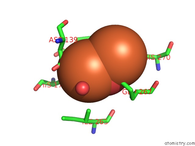
Mono view
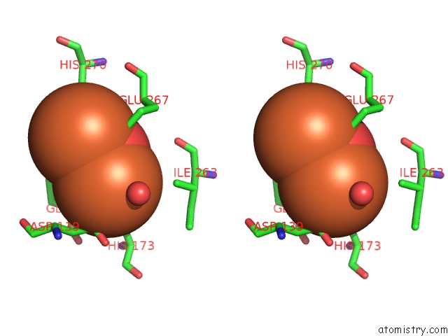
Stereo pair view
Mono view
Stereo pair view
A full contact list of Iron with other atoms in the Fe binding
site number 1 of Crystal Structure of Mouse Ribonucleotide Reductase Subunit R2 Under Oxidizing Conditions. A Fully Occupied Dinuclear Iron Cluster. within 5.0Å range:
|
Iron binding site 2 out of 2 in 1w68
Go back to
Iron binding site 2 out
of 2 in the Crystal Structure of Mouse Ribonucleotide Reductase Subunit R2 Under Oxidizing Conditions. A Fully Occupied Dinuclear Iron Cluster.
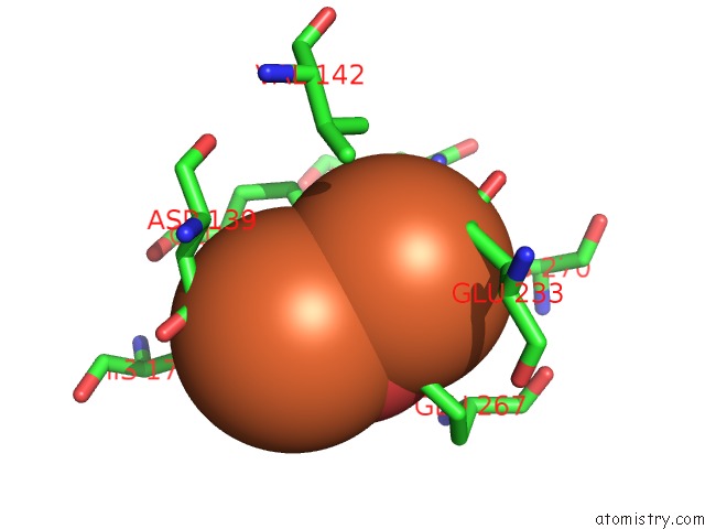
Mono view

Stereo pair view
Mono view
Stereo pair view
A full contact list of Iron with other atoms in the Fe binding
site number 2 of Crystal Structure of Mouse Ribonucleotide Reductase Subunit R2 Under Oxidizing Conditions. A Fully Occupied Dinuclear Iron Cluster. within 5.0Å range:
|
Reference:
K.R.Strand,
S.Karlsen,
M.Kolberg,
A.K.Rohr,
C.H.Gorbitz,
K.K.Andersson.
Crystal Structural Studies of Changes in the Native Dinuclear Iron Center of Ribonucleotide Reductase Protein R2 From Mouse J.Biol.Chem. V. 279 46794 2004.
ISSN: ISSN 0021-9258
PubMed: 15322079
DOI: 10.1074/JBC.M407346200
Page generated: Sat Aug 3 16:27:06 2024
ISSN: ISSN 0021-9258
PubMed: 15322079
DOI: 10.1074/JBC.M407346200
Last articles
Zn in 9J0NZn in 9J0O
Zn in 9J0P
Zn in 9FJX
Zn in 9EKB
Zn in 9C0F
Zn in 9CAH
Zn in 9CH0
Zn in 9CH3
Zn in 9CH1