Iron »
PDB 1wa6-1x71 »
1wov »
Iron in PDB 1wov: Crystal Structure of Heme Oxygenase-2 From Synechocystis Sp. Pcc 6803 in Complex with Heme
Enzymatic activity of Crystal Structure of Heme Oxygenase-2 From Synechocystis Sp. Pcc 6803 in Complex with Heme
All present enzymatic activity of Crystal Structure of Heme Oxygenase-2 From Synechocystis Sp. Pcc 6803 in Complex with Heme:
1.14.99.3;
1.14.99.3;
Protein crystallography data
The structure of Crystal Structure of Heme Oxygenase-2 From Synechocystis Sp. Pcc 6803 in Complex with Heme, PDB code: 1wov
was solved by
M.Sugishima,
Y.Hagiwara,
X.Zhang,
T.Yoshida,
C.T.Migita,
K.Fukuyama,
with X-Ray Crystallography technique. A brief refinement statistics is given in the table below:
| Resolution Low / High (Å) | 19.76 / 1.75 |
| Space group | P 1 21 1 |
| Cell size a, b, c (Å), α, β, γ (°) | 58.163, 74.585, 72.661, 90.00, 108.15, 90.00 |
| R / Rfree (%) | 19.3 / 25.4 |
Iron Binding Sites:
The binding sites of Iron atom in the Crystal Structure of Heme Oxygenase-2 From Synechocystis Sp. Pcc 6803 in Complex with Heme
(pdb code 1wov). This binding sites where shown within
5.0 Angstroms radius around Iron atom.
In total 2 binding sites of Iron where determined in the Crystal Structure of Heme Oxygenase-2 From Synechocystis Sp. Pcc 6803 in Complex with Heme, PDB code: 1wov:
Jump to Iron binding site number: 1; 2;
In total 2 binding sites of Iron where determined in the Crystal Structure of Heme Oxygenase-2 From Synechocystis Sp. Pcc 6803 in Complex with Heme, PDB code: 1wov:
Jump to Iron binding site number: 1; 2;
Iron binding site 1 out of 2 in 1wov
Go back to
Iron binding site 1 out
of 2 in the Crystal Structure of Heme Oxygenase-2 From Synechocystis Sp. Pcc 6803 in Complex with Heme
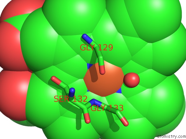
Mono view
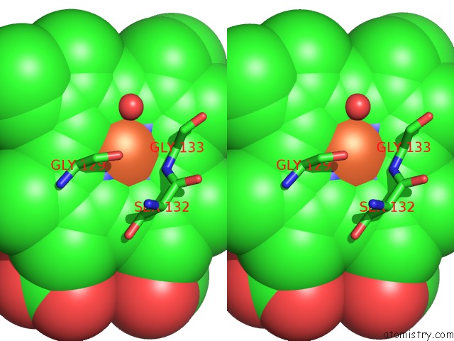
Stereo pair view
Mono view
Stereo pair view
A full contact list of Iron with other atoms in the Fe binding
site number 1 of Crystal Structure of Heme Oxygenase-2 From Synechocystis Sp. Pcc 6803 in Complex with Heme within 5.0Å range:
|
Iron binding site 2 out of 2 in 1wov
Go back to
Iron binding site 2 out
of 2 in the Crystal Structure of Heme Oxygenase-2 From Synechocystis Sp. Pcc 6803 in Complex with Heme
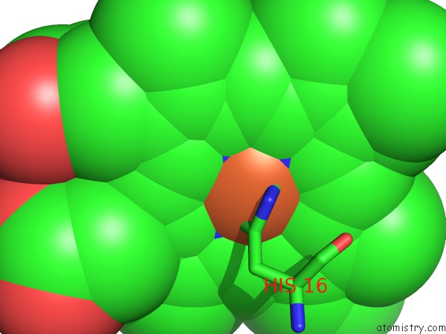
Mono view
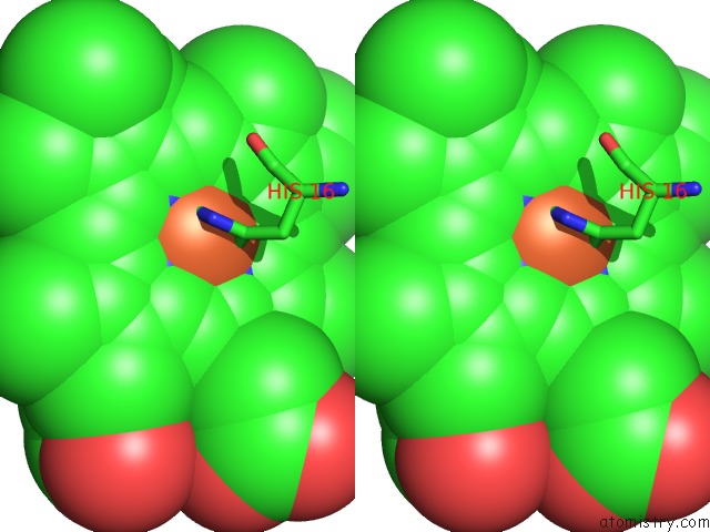
Stereo pair view
Mono view
Stereo pair view
A full contact list of Iron with other atoms in the Fe binding
site number 2 of Crystal Structure of Heme Oxygenase-2 From Synechocystis Sp. Pcc 6803 in Complex with Heme within 5.0Å range:
|
Reference:
M.Sugishima,
Y.Hagiwara,
X.Zhang,
T.Yoshida,
C.T.Migita,
K.Fukuyama.
Crystal Structure of Dimeric Heme Oxygenase-2 From Synechocystis Sp. Pcc 6803 in Complex with Heme. Biochemistry V. 44 4257 2005.
ISSN: ISSN 0006-2960
PubMed: 15766254
DOI: 10.1021/BI0480483
Page generated: Sat Aug 3 16:33:53 2024
ISSN: ISSN 0006-2960
PubMed: 15766254
DOI: 10.1021/BI0480483
Last articles
Zn in 9J0NZn in 9J0O
Zn in 9J0P
Zn in 9FJX
Zn in 9EKB
Zn in 9C0F
Zn in 9CAH
Zn in 9CH0
Zn in 9CH3
Zn in 9CH1