Iron »
PDB 1wa6-1x71 »
1wyg »
Iron in PDB 1wyg: Crystal Structure of A Rat Xanthine Dehydrogenase Triple Mutant (C535A, C992R and C1324S)
Enzymatic activity of Crystal Structure of A Rat Xanthine Dehydrogenase Triple Mutant (C535A, C992R and C1324S)
All present enzymatic activity of Crystal Structure of A Rat Xanthine Dehydrogenase Triple Mutant (C535A, C992R and C1324S):
1.1.1.204; 1.1.3.22;
1.1.1.204; 1.1.3.22;
Protein crystallography data
The structure of Crystal Structure of A Rat Xanthine Dehydrogenase Triple Mutant (C535A, C992R and C1324S), PDB code: 1wyg
was solved by
T.Nishino,
K.Okamoto,
Y.Kawaguchi,
H.Hori,
T.Matsumura,
B.T.Eger,
E.F.Pai,
T.Nishino,
with X-Ray Crystallography technique. A brief refinement statistics is given in the table below:
| Resolution Low / High (Å) | 25.00 / 2.60 |
| Space group | I 41 2 2 |
| Cell size a, b, c (Å), α, β, γ (°) | 134.253, 134.253, 523.315, 90.00, 90.00, 90.00 |
| R / Rfree (%) | 20.3 / 24.8 |
Other elements in 1wyg:
The structure of Crystal Structure of A Rat Xanthine Dehydrogenase Triple Mutant (C535A, C992R and C1324S) also contains other interesting chemical elements:
| Calcium | (Ca) | 1 atom |
Iron Binding Sites:
The binding sites of Iron atom in the Crystal Structure of A Rat Xanthine Dehydrogenase Triple Mutant (C535A, C992R and C1324S)
(pdb code 1wyg). This binding sites where shown within
5.0 Angstroms radius around Iron atom.
In total 4 binding sites of Iron where determined in the Crystal Structure of A Rat Xanthine Dehydrogenase Triple Mutant (C535A, C992R and C1324S), PDB code: 1wyg:
Jump to Iron binding site number: 1; 2; 3; 4;
In total 4 binding sites of Iron where determined in the Crystal Structure of A Rat Xanthine Dehydrogenase Triple Mutant (C535A, C992R and C1324S), PDB code: 1wyg:
Jump to Iron binding site number: 1; 2; 3; 4;
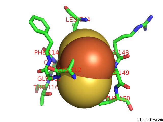
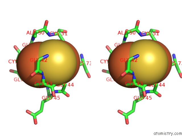
Iron binding site 1 out of 4 in 1wyg
Go back to
Iron binding site 1 out
of 4 in the Crystal Structure of A Rat Xanthine Dehydrogenase Triple Mutant (C535A, C992R and C1324S)
Mono view
Stereo pair view
Mono view
Stereo pair view
A full contact list of Iron with other atoms in the Fe binding
site number 1 of Crystal Structure of A Rat Xanthine Dehydrogenase Triple Mutant (C535A, C992R and C1324S) within 5.0Å range:
|
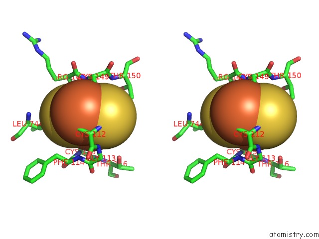
Iron binding site 2 out of 4 in 1wyg
Go back to
Iron binding site 2 out
of 4 in the Crystal Structure of A Rat Xanthine Dehydrogenase Triple Mutant (C535A, C992R and C1324S)
Mono view

Stereo pair view
Mono view
Stereo pair view
A full contact list of Iron with other atoms in the Fe binding
site number 2 of Crystal Structure of A Rat Xanthine Dehydrogenase Triple Mutant (C535A, C992R and C1324S) within 5.0Å range:
|
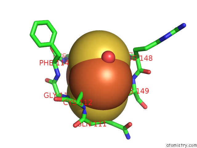
Iron binding site 3 out of 4 in 1wyg
Go back to
Iron binding site 3 out
of 4 in the Crystal Structure of A Rat Xanthine Dehydrogenase Triple Mutant (C535A, C992R and C1324S)

Mono view
Stereo pair view
Mono view
Stereo pair view
A full contact list of Iron with other atoms in the Fe binding
site number 3 of Crystal Structure of A Rat Xanthine Dehydrogenase Triple Mutant (C535A, C992R and C1324S) within 5.0Å range:
|
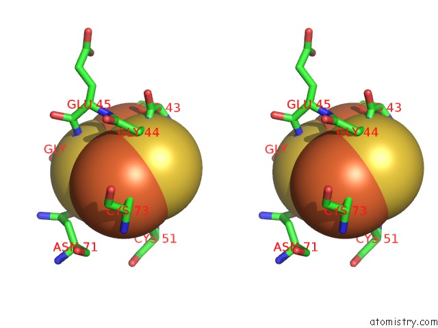
Iron binding site 4 out of 4 in 1wyg
Go back to
Iron binding site 4 out
of 4 in the Crystal Structure of A Rat Xanthine Dehydrogenase Triple Mutant (C535A, C992R and C1324S)

Mono view
Stereo pair view
Mono view
Stereo pair view
A full contact list of Iron with other atoms in the Fe binding
site number 4 of Crystal Structure of A Rat Xanthine Dehydrogenase Triple Mutant (C535A, C992R and C1324S) within 5.0Å range:
|
Reference:
T.Nishino,
K.Okamoto,
Y.Kawaguchi,
H.Hori,
T.Matsumura,
B.T.Eger,
E.F.Pai,
T.Nishino.
Mechanism of the Conversion of Xanthine Dehydrogenase to Xanthine Oxidase: Identification of the Two Cysteine Disulfide Bonds and Crystal Structure of A Non-Convertible Rat Liver Xanthine Dehydrogenase Mutant J.Biol.Chem. V. 280 24888 2005.
ISSN: ISSN 0021-9258
PubMed: 15878860
DOI: 10.1074/JBC.M501830200
Page generated: Sat Aug 3 16:40:38 2024
ISSN: ISSN 0021-9258
PubMed: 15878860
DOI: 10.1074/JBC.M501830200
Last articles
Zn in 9J0NZn in 9J0O
Zn in 9J0P
Zn in 9FJX
Zn in 9EKB
Zn in 9C0F
Zn in 9CAH
Zn in 9CH0
Zn in 9CH3
Zn in 9CH1