Iron »
PDB 1wa6-1x71 »
1x46 »
Iron in PDB 1x46: Crystal Structure of A Hemoglobin Component (Ta-VII) From Tokunagayusurika Akamusi
Protein crystallography data
The structure of Crystal Structure of A Hemoglobin Component (Ta-VII) From Tokunagayusurika Akamusi, PDB code: 1x46
was solved by
T.Kuwada,
T.Hasegawa,
S.Sato,
I.Sato,
K.Ishikawa,
T.Takagi,
F.Shishikura,
with X-Ray Crystallography technique. A brief refinement statistics is given in the table below:
| Resolution Low / High (Å) | 20.21 / 1.50 |
| Space group | C 2 2 21 |
| Cell size a, b, c (Å), α, β, γ (°) | 42.010, 69.110, 99.650, 90.00, 90.00, 90.00 |
| R / Rfree (%) | n/a / n/a |
Iron Binding Sites:
The binding sites of Iron atom in the Crystal Structure of A Hemoglobin Component (Ta-VII) From Tokunagayusurika Akamusi
(pdb code 1x46). This binding sites where shown within
5.0 Angstroms radius around Iron atom.
In total only one binding site of Iron was determined in the Crystal Structure of A Hemoglobin Component (Ta-VII) From Tokunagayusurika Akamusi, PDB code: 1x46:
In total only one binding site of Iron was determined in the Crystal Structure of A Hemoglobin Component (Ta-VII) From Tokunagayusurika Akamusi, PDB code: 1x46:
Iron binding site 1 out of 1 in 1x46
Go back to
Iron binding site 1 out
of 1 in the Crystal Structure of A Hemoglobin Component (Ta-VII) From Tokunagayusurika Akamusi

Mono view
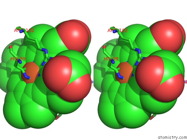
Stereo pair view
Mono view
Stereo pair view
A full contact list of Iron with other atoms in the Fe binding
site number 1 of Crystal Structure of A Hemoglobin Component (Ta-VII) From Tokunagayusurika Akamusi within 5.0Å range:
|
Reference:
T.Kuwada,
T.Hasegawa,
S.Sato,
I.Sato,
K.Ishikawa,
T.Takagi,
F.Shishikura.
Crystal Structures of Two Hemoglobin Components From the Midge Larva Propsilocerus Akamusi (Orthocladiinae, Diptera). Gene V. 398 29 2007.
ISSN: ISSN 0378-1119
PubMed: 17590288
DOI: 10.1016/J.GENE.2007.02.049
Page generated: Sat Aug 3 16:46:27 2024
ISSN: ISSN 0378-1119
PubMed: 17590288
DOI: 10.1016/J.GENE.2007.02.049
Last articles
Zn in 9J0NZn in 9J0O
Zn in 9J0P
Zn in 9FJX
Zn in 9EKB
Zn in 9C0F
Zn in 9CAH
Zn in 9CH0
Zn in 9CH3
Zn in 9CH1