Iron »
PDB 1x89-1xvc »
1xbn »
Iron in PDB 1xbn: Crystal Structure of A Bacterial Nitric Oxide Sensor: An Ortholog of Mammalian Soluble Guanylate Cyclase Heme Domain
Protein crystallography data
The structure of Crystal Structure of A Bacterial Nitric Oxide Sensor: An Ortholog of Mammalian Soluble Guanylate Cyclase Heme Domain, PDB code: 1xbn
was solved by
P.Nioche,
C.S.Raman,
with X-Ray Crystallography technique. A brief refinement statistics is given in the table below:
| Resolution Low / High (Å) | 84.51 / 2.50 |
| Space group | P 42 3 2 |
| Cell size a, b, c (Å), α, β, γ (°) | 120.607, 120.607, 120.607, 90.00, 90.00, 90.00 |
| R / Rfree (%) | 26.3 / 29.1 |
Iron Binding Sites:
The binding sites of Iron atom in the Crystal Structure of A Bacterial Nitric Oxide Sensor: An Ortholog of Mammalian Soluble Guanylate Cyclase Heme Domain
(pdb code 1xbn). This binding sites where shown within
5.0 Angstroms radius around Iron atom.
In total only one binding site of Iron was determined in the Crystal Structure of A Bacterial Nitric Oxide Sensor: An Ortholog of Mammalian Soluble Guanylate Cyclase Heme Domain, PDB code: 1xbn:
In total only one binding site of Iron was determined in the Crystal Structure of A Bacterial Nitric Oxide Sensor: An Ortholog of Mammalian Soluble Guanylate Cyclase Heme Domain, PDB code: 1xbn:
Iron binding site 1 out of 1 in 1xbn
Go back to
Iron binding site 1 out
of 1 in the Crystal Structure of A Bacterial Nitric Oxide Sensor: An Ortholog of Mammalian Soluble Guanylate Cyclase Heme Domain
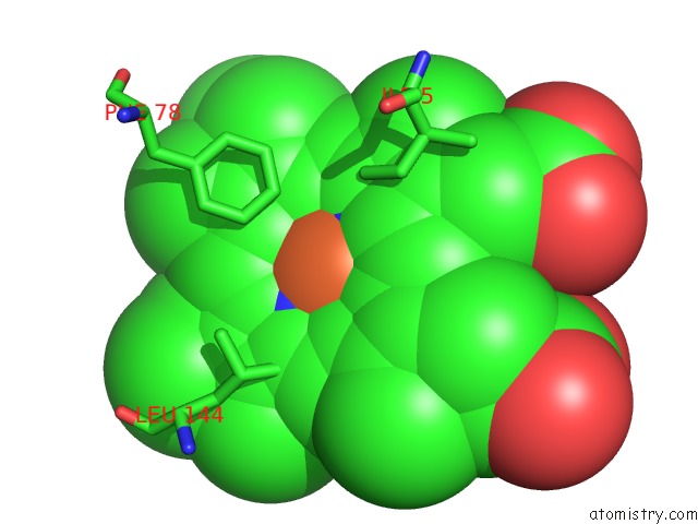
Mono view
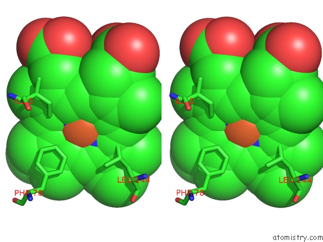
Stereo pair view
Mono view
Stereo pair view
A full contact list of Iron with other atoms in the Fe binding
site number 1 of Crystal Structure of A Bacterial Nitric Oxide Sensor: An Ortholog of Mammalian Soluble Guanylate Cyclase Heme Domain within 5.0Å range:
|
Reference:
P.Nioche,
V.Berka,
J.Vipond,
N.Minton,
A.-L.Tsai,
C.S.Raman.
Femtomolar Sensitivity of A No Sensor From Clostridium Botulinum Science V. 306 1550 2004.
ISSN: ISSN 0036-8075
PubMed: 15472039
DOI: 10.1126/SCIENCE.1103596
Page generated: Sat Aug 3 16:51:02 2024
ISSN: ISSN 0036-8075
PubMed: 15472039
DOI: 10.1126/SCIENCE.1103596
Last articles
Zn in 9MJ5Zn in 9HNW
Zn in 9G0L
Zn in 9FNE
Zn in 9DZN
Zn in 9E0I
Zn in 9D32
Zn in 9DAK
Zn in 8ZXC
Zn in 8ZUF