Iron »
PDB 1x89-1xvc »
1xdb »
Iron in PDB 1xdb: Crystal Structure of the Nitrogenase Fe Protein ASP129GLU
Enzymatic activity of Crystal Structure of the Nitrogenase Fe Protein ASP129GLU
All present enzymatic activity of Crystal Structure of the Nitrogenase Fe Protein ASP129GLU:
1.18.6.1;
1.18.6.1;
Protein crystallography data
The structure of Crystal Structure of the Nitrogenase Fe Protein ASP129GLU, PDB code: 1xdb
was solved by
S.B.Jang,
M.S.Jeong,
L.C.Seefeldt,
J.W.Peters,
with X-Ray Crystallography technique. A brief refinement statistics is given in the table below:
| Resolution Low / High (Å) | 20.00 / 2.80 |
| Space group | P 1 21 1 |
| Cell size a, b, c (Å), α, β, γ (°) | 57.574, 92.138, 63.934, 90.00, 100.80, 90.00 |
| R / Rfree (%) | 22.7 / 29.9 |
Iron Binding Sites:
The binding sites of Iron atom in the Crystal Structure of the Nitrogenase Fe Protein ASP129GLU
(pdb code 1xdb). This binding sites where shown within
5.0 Angstroms radius around Iron atom.
In total 4 binding sites of Iron where determined in the Crystal Structure of the Nitrogenase Fe Protein ASP129GLU, PDB code: 1xdb:
Jump to Iron binding site number: 1; 2; 3; 4;
In total 4 binding sites of Iron where determined in the Crystal Structure of the Nitrogenase Fe Protein ASP129GLU, PDB code: 1xdb:
Jump to Iron binding site number: 1; 2; 3; 4;
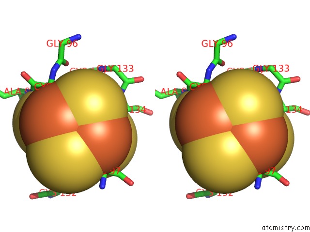
Iron binding site 1 out of 4 in 1xdb
Go back to
Iron binding site 1 out
of 4 in the Crystal Structure of the Nitrogenase Fe Protein ASP129GLU
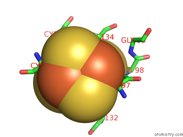
Mono view
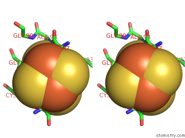
Stereo pair view
Mono view
Stereo pair view
A full contact list of Iron with other atoms in the Fe binding
site number 1 of Crystal Structure of the Nitrogenase Fe Protein ASP129GLU within 5.0Å range:
|
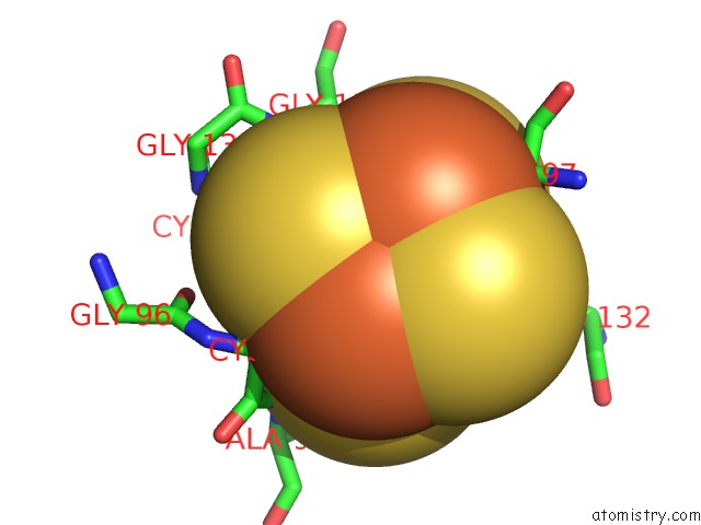
Iron binding site 2 out of 4 in 1xdb
Go back to
Iron binding site 2 out
of 4 in the Crystal Structure of the Nitrogenase Fe Protein ASP129GLU
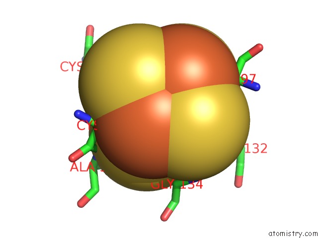
Mono view

Stereo pair view
Mono view
Stereo pair view
A full contact list of Iron with other atoms in the Fe binding
site number 2 of Crystal Structure of the Nitrogenase Fe Protein ASP129GLU within 5.0Å range:
|
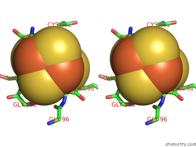
Iron binding site 3 out of 4 in 1xdb
Go back to
Iron binding site 3 out
of 4 in the Crystal Structure of the Nitrogenase Fe Protein ASP129GLU
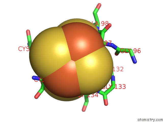
Mono view
Stereo pair view
Mono view
Stereo pair view
A full contact list of Iron with other atoms in the Fe binding
site number 3 of Crystal Structure of the Nitrogenase Fe Protein ASP129GLU within 5.0Å range:
|
Iron binding site 4 out of 4 in 1xdb
Go back to
Iron binding site 4 out
of 4 in the Crystal Structure of the Nitrogenase Fe Protein ASP129GLU
Mono view
Stereo pair view
Mono view
Stereo pair view
A full contact list of Iron with other atoms in the Fe binding
site number 4 of Crystal Structure of the Nitrogenase Fe Protein ASP129GLU within 5.0Å range:
|
Reference:
S.B.Jang,
M.S.Jeong,
L.C.Seefeldt,
J.W.Peters.
Structural and Biochemical Implications of Single Amino Acid Substitutions in the Nucleotide-Dependent Switch Regions of the Nitrogenase Fe Protein From Azotobacter Vinelandii J.Biol.Inorg.Chem. V. 9 1028 2004.
ISSN: ISSN 0949-8257
PubMed: 15549494
DOI: 10.1007/S00775-004-0605-5
Page generated: Sat Aug 3 16:52:14 2024
ISSN: ISSN 0949-8257
PubMed: 15549494
DOI: 10.1007/S00775-004-0605-5
Last articles
Zn in 9MJ5Zn in 9HNW
Zn in 9G0L
Zn in 9FNE
Zn in 9DZN
Zn in 9E0I
Zn in 9D32
Zn in 9DAK
Zn in 8ZXC
Zn in 8ZUF