Iron »
PDB 1x89-1xvc »
1xen »
Iron in PDB 1xen: High Resolution Crystal Structure of Escherichia Coli Iron- Peptide Deformylase Bound to Formate
Enzymatic activity of High Resolution Crystal Structure of Escherichia Coli Iron- Peptide Deformylase Bound to Formate
All present enzymatic activity of High Resolution Crystal Structure of Escherichia Coli Iron- Peptide Deformylase Bound to Formate:
3.5.1.88;
3.5.1.88;
Protein crystallography data
The structure of High Resolution Crystal Structure of Escherichia Coli Iron- Peptide Deformylase Bound to Formate, PDB code: 1xen
was solved by
R.Jain,
B.Hao,
R.-P.Liu,
M.K.Chan,
with X-Ray Crystallography technique. A brief refinement statistics is given in the table below:
| Resolution Low / High (Å) | 19.61 / 1.85 |
| Space group | P 61 2 2 |
| Cell size a, b, c (Å), α, β, γ (°) | 54.820, 54.820, 224.580, 90.00, 90.00, 120.00 |
| R / Rfree (%) | 17.3 / 21.8 |
Iron Binding Sites:
The binding sites of Iron atom in the High Resolution Crystal Structure of Escherichia Coli Iron- Peptide Deformylase Bound to Formate
(pdb code 1xen). This binding sites where shown within
5.0 Angstroms radius around Iron atom.
In total only one binding site of Iron was determined in the High Resolution Crystal Structure of Escherichia Coli Iron- Peptide Deformylase Bound to Formate, PDB code: 1xen:
In total only one binding site of Iron was determined in the High Resolution Crystal Structure of Escherichia Coli Iron- Peptide Deformylase Bound to Formate, PDB code: 1xen:
Iron binding site 1 out of 1 in 1xen
Go back to
Iron binding site 1 out
of 1 in the High Resolution Crystal Structure of Escherichia Coli Iron- Peptide Deformylase Bound to Formate
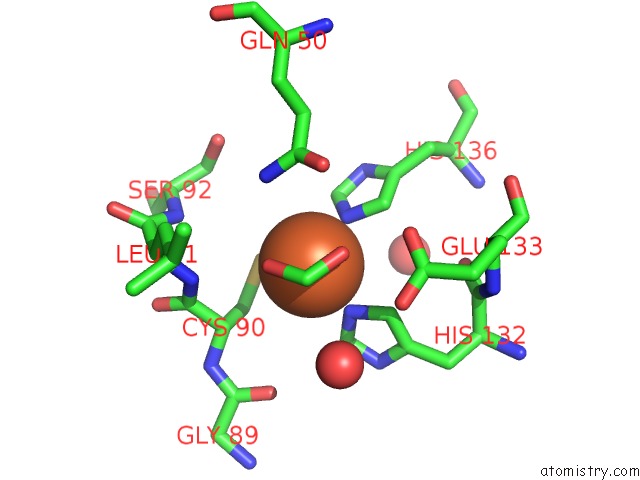
Mono view
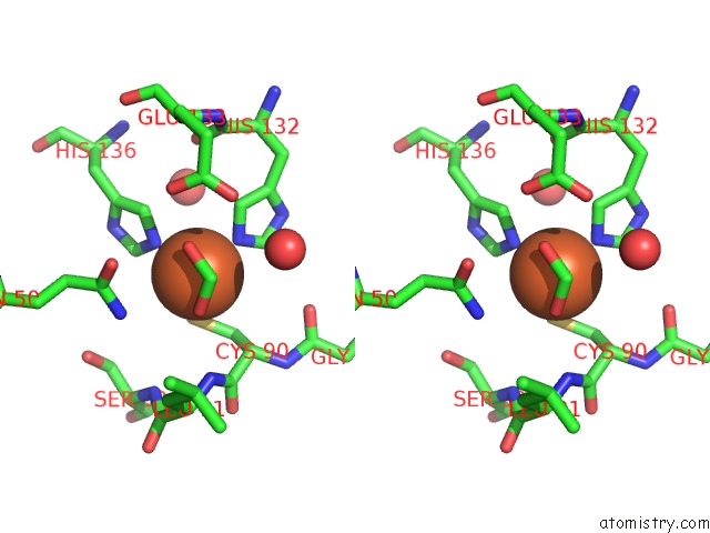
Stereo pair view
Mono view
Stereo pair view
A full contact list of Iron with other atoms in the Fe binding
site number 1 of High Resolution Crystal Structure of Escherichia Coli Iron- Peptide Deformylase Bound to Formate within 5.0Å range:
|
Reference:
R.Jain,
B.Hao,
R.-P.Liu,
M.K.Chan.
Structures of E. Coli Peptide Deformylase Bound to Formate: Insight Into the Preference For FE2+ Over ZN2+ As the Active Site Metal J.Am.Chem.Soc. V. 127 4558 2005.
ISSN: ISSN 0002-7863
PubMed: 15796505
DOI: 10.1021/JA0503074
Page generated: Sat Aug 3 16:52:16 2024
ISSN: ISSN 0002-7863
PubMed: 15796505
DOI: 10.1021/JA0503074
Last articles
Zn in 9MJ5Zn in 9HNW
Zn in 9G0L
Zn in 9FNE
Zn in 9DZN
Zn in 9E0I
Zn in 9D32
Zn in 9DAK
Zn in 8ZXC
Zn in 8ZUF