Iron »
PDB 1x89-1xvc »
1xk0 »
Iron in PDB 1xk0: Crystal Structures of the G139A, G139A-No and G143H Mutants of Human Heme Oxygenase-1
Enzymatic activity of Crystal Structures of the G139A, G139A-No and G143H Mutants of Human Heme Oxygenase-1
All present enzymatic activity of Crystal Structures of the G139A, G139A-No and G143H Mutants of Human Heme Oxygenase-1:
1.14.99.3;
1.14.99.3;
Protein crystallography data
The structure of Crystal Structures of the G139A, G139A-No and G143H Mutants of Human Heme Oxygenase-1, PDB code: 1xk0
was solved by
L.Lad,
A.Koshkin,
P.R.Ortiz De Montellano,
T.L.Poulos,
with X-Ray Crystallography technique. A brief refinement statistics is given in the table below:
| Resolution Low / High (Å) | 50.00 / 2.18 |
| Space group | P 21 21 21 |
| Cell size a, b, c (Å), α, β, γ (°) | 54.060, 75.968, 105.299, 90.00, 90.00, 90.00 |
| R / Rfree (%) | 22.4 / 26.6 |
Iron Binding Sites:
The binding sites of Iron atom in the Crystal Structures of the G139A, G139A-No and G143H Mutants of Human Heme Oxygenase-1
(pdb code 1xk0). This binding sites where shown within
5.0 Angstroms radius around Iron atom.
In total 2 binding sites of Iron where determined in the Crystal Structures of the G139A, G139A-No and G143H Mutants of Human Heme Oxygenase-1, PDB code: 1xk0:
Jump to Iron binding site number: 1; 2;
In total 2 binding sites of Iron where determined in the Crystal Structures of the G139A, G139A-No and G143H Mutants of Human Heme Oxygenase-1, PDB code: 1xk0:
Jump to Iron binding site number: 1; 2;
Iron binding site 1 out of 2 in 1xk0
Go back to
Iron binding site 1 out
of 2 in the Crystal Structures of the G139A, G139A-No and G143H Mutants of Human Heme Oxygenase-1
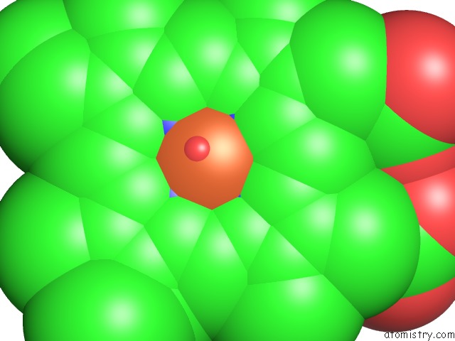
Mono view
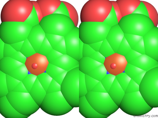
Stereo pair view
Mono view
Stereo pair view
A full contact list of Iron with other atoms in the Fe binding
site number 1 of Crystal Structures of the G139A, G139A-No and G143H Mutants of Human Heme Oxygenase-1 within 5.0Å range:
|
Iron binding site 2 out of 2 in 1xk0
Go back to
Iron binding site 2 out
of 2 in the Crystal Structures of the G139A, G139A-No and G143H Mutants of Human Heme Oxygenase-1
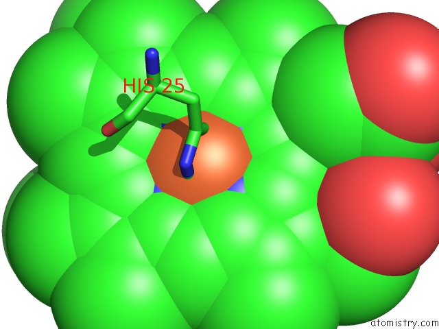
Mono view
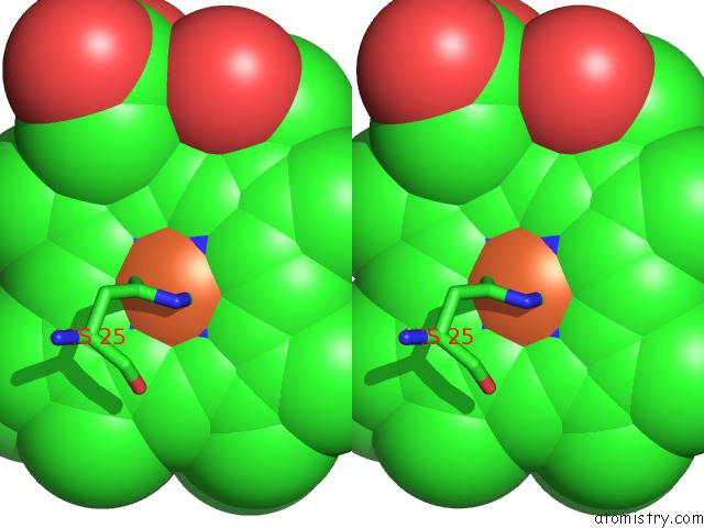
Stereo pair view
Mono view
Stereo pair view
A full contact list of Iron with other atoms in the Fe binding
site number 2 of Crystal Structures of the G139A, G139A-No and G143H Mutants of Human Heme Oxygenase-1 within 5.0Å range:
|
Reference:
L.Lad,
A.Koshkin,
P.R.Ortiz De Montellano,
T.L.Poulos.
Crystal Structures of the G139A, G139A-No and G143H Mutants of Human Heme Oxygenase-1. A Finely Tuned Hydrogen-Bonding Network Controls Oxygenase Versus Peroxidase Activity. J.Biol.Inorg.Chem. V. 10 138 2005.
ISSN: ISSN 0949-8257
PubMed: 15690204
DOI: 10.1007/S00775-004-0620-6
Page generated: Sat Aug 3 16:56:03 2024
ISSN: ISSN 0949-8257
PubMed: 15690204
DOI: 10.1007/S00775-004-0620-6
Last articles
Zn in 9MJ5Zn in 9HNW
Zn in 9G0L
Zn in 9FNE
Zn in 9DZN
Zn in 9E0I
Zn in 9D32
Zn in 9DAK
Zn in 8ZXC
Zn in 8ZUF