Iron »
PDB 1x89-1xvc »
1xqd »
Iron in PDB 1xqd: Crystal Structure of P450NOR Complexed with 3- Pyridinealdehyde Adenine Dinucleotide
Enzymatic activity of Crystal Structure of P450NOR Complexed with 3- Pyridinealdehyde Adenine Dinucleotide
All present enzymatic activity of Crystal Structure of P450NOR Complexed with 3- Pyridinealdehyde Adenine Dinucleotide:
1.7.99.7;
1.7.99.7;
Protein crystallography data
The structure of Crystal Structure of P450NOR Complexed with 3- Pyridinealdehyde Adenine Dinucleotide, PDB code: 1xqd
was solved by
R.Oshima,
S.Fushinobu,
N.Takaya,
F.Su,
T.Wakagi,
H.Shoun,
with X-Ray Crystallography technique. A brief refinement statistics is given in the table below:
| Resolution Low / High (Å) | 19.54 / 1.80 |
| Space group | P 21 21 21 |
| Cell size a, b, c (Å), α, β, γ (°) | 43.296, 56.121, 163.262, 90.00, 90.00, 90.00 |
| R / Rfree (%) | 20.6 / 24.3 |
Iron Binding Sites:
The binding sites of Iron atom in the Crystal Structure of P450NOR Complexed with 3- Pyridinealdehyde Adenine Dinucleotide
(pdb code 1xqd). This binding sites where shown within
5.0 Angstroms radius around Iron atom.
In total only one binding site of Iron was determined in the Crystal Structure of P450NOR Complexed with 3- Pyridinealdehyde Adenine Dinucleotide, PDB code: 1xqd:
In total only one binding site of Iron was determined in the Crystal Structure of P450NOR Complexed with 3- Pyridinealdehyde Adenine Dinucleotide, PDB code: 1xqd:
Iron binding site 1 out of 1 in 1xqd
Go back to
Iron binding site 1 out
of 1 in the Crystal Structure of P450NOR Complexed with 3- Pyridinealdehyde Adenine Dinucleotide
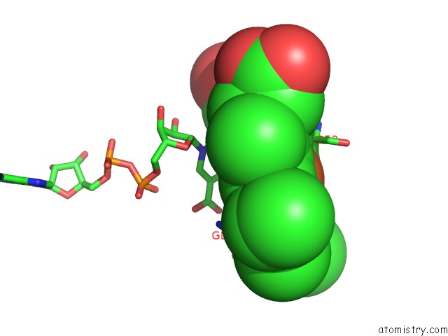
Mono view
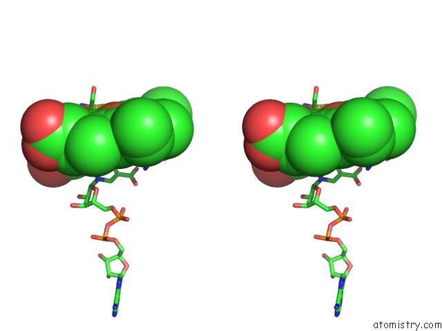
Stereo pair view
Mono view
Stereo pair view
A full contact list of Iron with other atoms in the Fe binding
site number 1 of Crystal Structure of P450NOR Complexed with 3- Pyridinealdehyde Adenine Dinucleotide within 5.0Å range:
|
Reference:
R.Oshima,
S.Fushinobu,
F.Su,
L.Zhang,
N.Takaya,
H.Shoun.
Structural Evidence For Direct Hydride Transfer From Nadh to Cytochrome P450NOR J.Mol.Biol. V. 342 207 2004.
ISSN: ISSN 0022-2836
PubMed: 15313618
DOI: 10.1016/J.JMB.2004.07.009
Page generated: Sat Aug 3 16:59:26 2024
ISSN: ISSN 0022-2836
PubMed: 15313618
DOI: 10.1016/J.JMB.2004.07.009
Last articles
Zn in 9MJ5Zn in 9HNW
Zn in 9G0L
Zn in 9FNE
Zn in 9DZN
Zn in 9E0I
Zn in 9D32
Zn in 9DAK
Zn in 8ZXC
Zn in 8ZUF