Iron »
PDB 1x89-1xvc »
1xsm »
Iron in PDB 1xsm: Protein R2 of Ribonucleotide Reductase From Mouse
Enzymatic activity of Protein R2 of Ribonucleotide Reductase From Mouse
All present enzymatic activity of Protein R2 of Ribonucleotide Reductase From Mouse:
1.17.4.1;
1.17.4.1;
Protein crystallography data
The structure of Protein R2 of Ribonucleotide Reductase From Mouse, PDB code: 1xsm
was solved by
B.Kauppi,
B.N.Nielsen,
S.Ramaswamy,
I.Kjoller-Larsen,
M.Thelander,
L.Thelander,
H.Eklund,
with X-Ray Crystallography technique. A brief refinement statistics is given in the table below:
| Resolution Low / High (Å) | 25.00 / 2.30 |
| Space group | C 2 2 21 |
| Cell size a, b, c (Å), α, β, γ (°) | 77.078, 108.932, 92.900, 90.00, 90.00, 90.00 |
| R / Rfree (%) | 19.1 / 25 |
Iron Binding Sites:
The binding sites of Iron atom in the Protein R2 of Ribonucleotide Reductase From Mouse
(pdb code 1xsm). This binding sites where shown within
5.0 Angstroms radius around Iron atom.
In total only one binding site of Iron was determined in the Protein R2 of Ribonucleotide Reductase From Mouse, PDB code: 1xsm:
In total only one binding site of Iron was determined in the Protein R2 of Ribonucleotide Reductase From Mouse, PDB code: 1xsm:
Iron binding site 1 out of 1 in 1xsm
Go back to
Iron binding site 1 out
of 1 in the Protein R2 of Ribonucleotide Reductase From Mouse
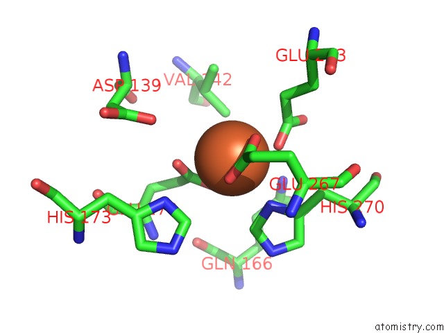
Mono view
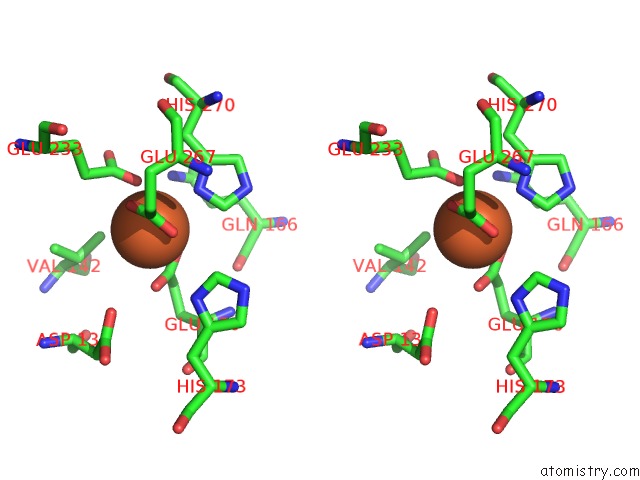
Stereo pair view
Mono view
Stereo pair view
A full contact list of Iron with other atoms in the Fe binding
site number 1 of Protein R2 of Ribonucleotide Reductase From Mouse within 5.0Å range:
|
Reference:
B.Kauppi,
B.B.Nielsen,
S.Ramaswamy,
I.K.Larsen,
M.Thelander,
L.Thelander,
H.Eklund.
The Three-Dimensional Structure of Mammalian Ribonucleotide Reductase Protein R2 Reveals A More-Accessible Iron-Radical Site Than Escherichia Coli R2. J.Mol.Biol. V. 262 706 1996.
ISSN: ISSN 0022-2836
PubMed: 8876648
DOI: 10.1006/JMBI.1996.0546
Page generated: Sat Aug 3 17:00:15 2024
ISSN: ISSN 0022-2836
PubMed: 8876648
DOI: 10.1006/JMBI.1996.0546
Last articles
Zn in 9MJ5Zn in 9HNW
Zn in 9G0L
Zn in 9FNE
Zn in 9DZN
Zn in 9E0I
Zn in 9D32
Zn in 9DAK
Zn in 8ZXC
Zn in 8ZUF