Iron »
PDB 1x89-1xvc »
1xu5 »
Iron in PDB 1xu5: Soluble Methane Monooxygenase Hydroxylase-Phenol Soaked
Enzymatic activity of Soluble Methane Monooxygenase Hydroxylase-Phenol Soaked
All present enzymatic activity of Soluble Methane Monooxygenase Hydroxylase-Phenol Soaked:
1.14.13.25;
1.14.13.25;
Protein crystallography data
The structure of Soluble Methane Monooxygenase Hydroxylase-Phenol Soaked, PDB code: 1xu5
was solved by
M.H.Sazinsky,
S.J.Lippard,
with X-Ray Crystallography technique. A brief refinement statistics is given in the table below:
| Resolution Low / High (Å) | 29.94 / 1.96 |
| Space group | P 21 21 21 |
| Cell size a, b, c (Å), α, β, γ (°) | 71.185, 171.558, 221.097, 90.00, 90.00, 90.00 |
| R / Rfree (%) | 20 / 22.8 |
Iron Binding Sites:
The binding sites of Iron atom in the Soluble Methane Monooxygenase Hydroxylase-Phenol Soaked
(pdb code 1xu5). This binding sites where shown within
5.0 Angstroms radius around Iron atom.
In total 4 binding sites of Iron where determined in the Soluble Methane Monooxygenase Hydroxylase-Phenol Soaked, PDB code: 1xu5:
Jump to Iron binding site number: 1; 2; 3; 4;
In total 4 binding sites of Iron where determined in the Soluble Methane Monooxygenase Hydroxylase-Phenol Soaked, PDB code: 1xu5:
Jump to Iron binding site number: 1; 2; 3; 4;
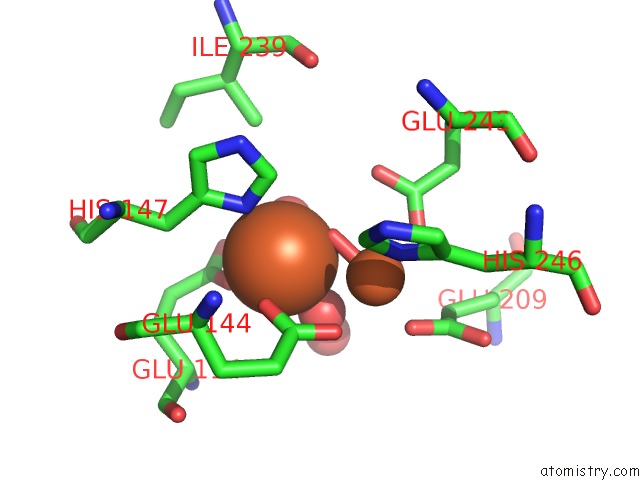
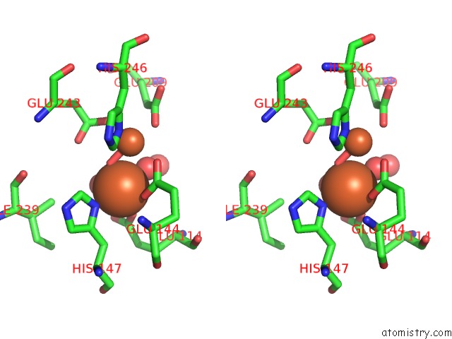
Iron binding site 1 out of 4 in 1xu5
Go back to
Iron binding site 1 out
of 4 in the Soluble Methane Monooxygenase Hydroxylase-Phenol Soaked
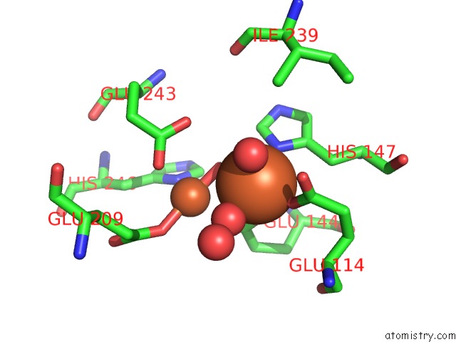
Mono view
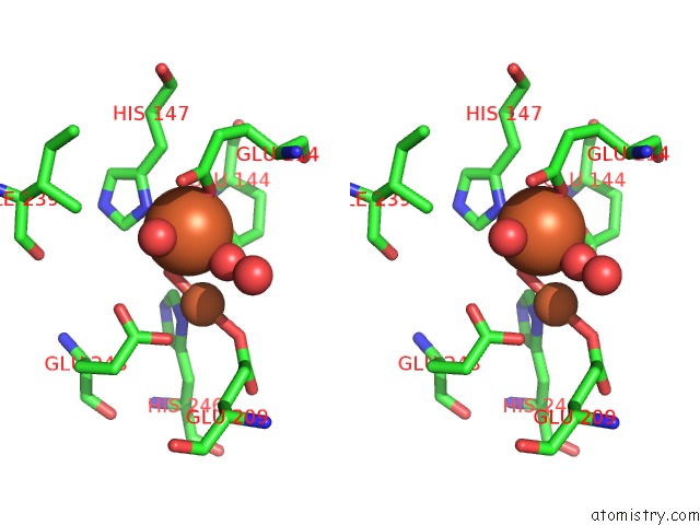
Stereo pair view
Mono view
Stereo pair view
A full contact list of Iron with other atoms in the Fe binding
site number 1 of Soluble Methane Monooxygenase Hydroxylase-Phenol Soaked within 5.0Å range:
|
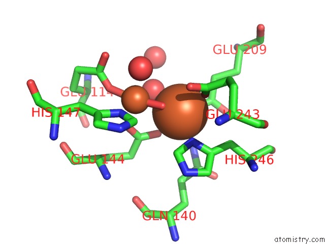
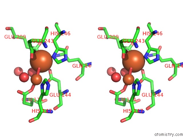
Iron binding site 2 out of 4 in 1xu5
Go back to
Iron binding site 2 out
of 4 in the Soluble Methane Monooxygenase Hydroxylase-Phenol Soaked
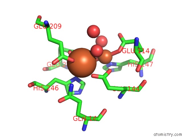
Mono view
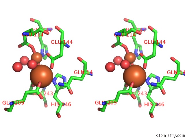
Stereo pair view
Mono view
Stereo pair view
A full contact list of Iron with other atoms in the Fe binding
site number 2 of Soluble Methane Monooxygenase Hydroxylase-Phenol Soaked within 5.0Å range:
|
Iron binding site 3 out of 4 in 1xu5
Go back to
Iron binding site 3 out
of 4 in the Soluble Methane Monooxygenase Hydroxylase-Phenol Soaked
Mono view
Stereo pair view
Mono view
Stereo pair view
A full contact list of Iron with other atoms in the Fe binding
site number 3 of Soluble Methane Monooxygenase Hydroxylase-Phenol Soaked within 5.0Å range:
|
Iron binding site 4 out of 4 in 1xu5
Go back to
Iron binding site 4 out
of 4 in the Soluble Methane Monooxygenase Hydroxylase-Phenol Soaked
Mono view
Stereo pair view
Mono view
Stereo pair view
A full contact list of Iron with other atoms in the Fe binding
site number 4 of Soluble Methane Monooxygenase Hydroxylase-Phenol Soaked within 5.0Å range:
|
Reference:
M.H.Sazinsky,
S.J.Lippard.
Product Bound Structures of the Soluble Methane Monooxygenase Hydroxylase From Methylococcus Capsulatus (Bath): Protein Motion in the Alpha-Subunit J.Am.Chem.Soc. V. 127 5814 2005.
ISSN: ISSN 0002-7863
PubMed: 15839679
DOI: 10.1021/JA044099B
Page generated: Sat Aug 3 17:00:37 2024
ISSN: ISSN 0002-7863
PubMed: 15839679
DOI: 10.1021/JA044099B
Last articles
F in 4LY6F in 4MM8
F in 4MIH
F in 4MM4
F in 4MK0
F in 4MIG
F in 4MH0
F in 4MDD
F in 4MDB
F in 4MC9