Iron »
PDB 1xvd-1y4q »
1y0a »
Iron in PDB 1y0a: T-to-Thigh Quaternary Transitions in Human Hemoglobin: ALPHAY140A Deoxy Low-Salt
Protein crystallography data
The structure of T-to-Thigh Quaternary Transitions in Human Hemoglobin: ALPHAY140A Deoxy Low-Salt, PDB code: 1y0a
was solved by
J.S.Kavanaugh,
P.H.Rogers,
A.Arnone,
with X-Ray Crystallography technique. A brief refinement statistics is given in the table below:
| Resolution Low / High (Å) | 10.00 / 2.22 |
| Space group | P 21 21 2 |
| Cell size a, b, c (Å), α, β, γ (°) | 96.800, 99.200, 65.800, 90.00, 90.00, 90.00 |
| R / Rfree (%) | 17.5 / 23.7 |
Iron Binding Sites:
The binding sites of Iron atom in the T-to-Thigh Quaternary Transitions in Human Hemoglobin: ALPHAY140A Deoxy Low-Salt
(pdb code 1y0a). This binding sites where shown within
5.0 Angstroms radius around Iron atom.
In total 4 binding sites of Iron where determined in the T-to-Thigh Quaternary Transitions in Human Hemoglobin: ALPHAY140A Deoxy Low-Salt, PDB code: 1y0a:
Jump to Iron binding site number: 1; 2; 3; 4;
In total 4 binding sites of Iron where determined in the T-to-Thigh Quaternary Transitions in Human Hemoglobin: ALPHAY140A Deoxy Low-Salt, PDB code: 1y0a:
Jump to Iron binding site number: 1; 2; 3; 4;
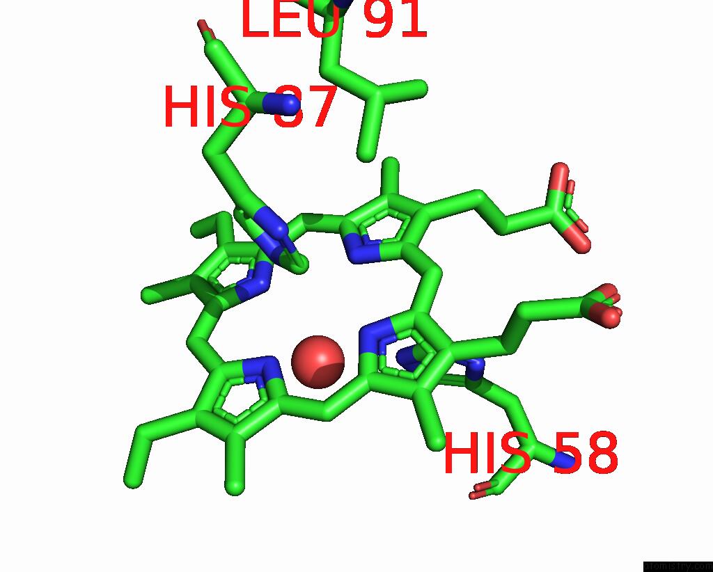
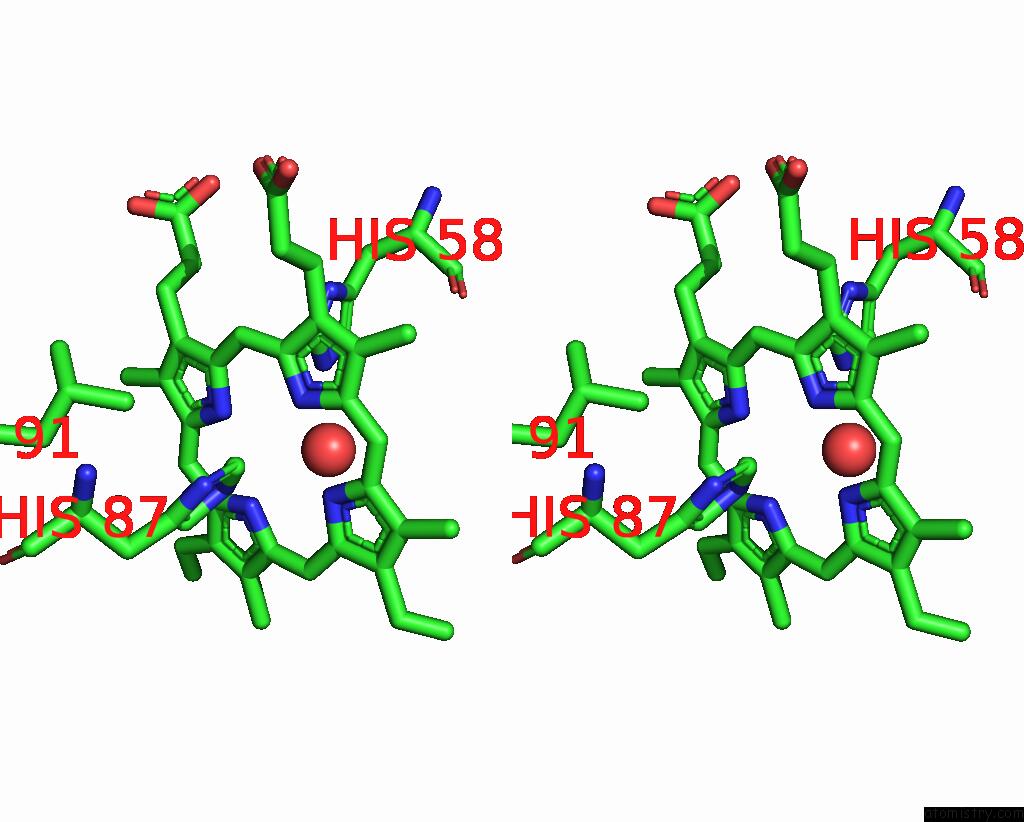
Iron binding site 1 out of 4 in 1y0a
Go back to
Iron binding site 1 out
of 4 in the T-to-Thigh Quaternary Transitions in Human Hemoglobin: ALPHAY140A Deoxy Low-Salt
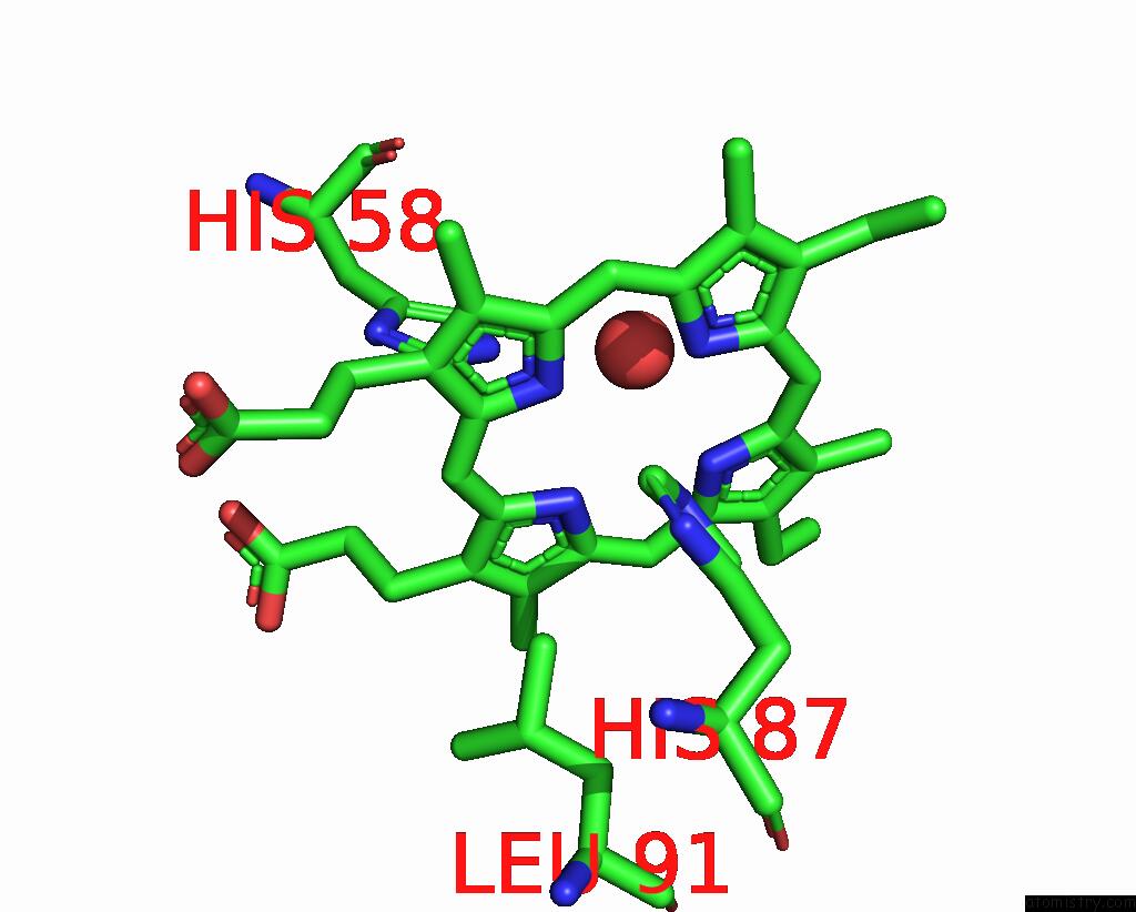
Mono view
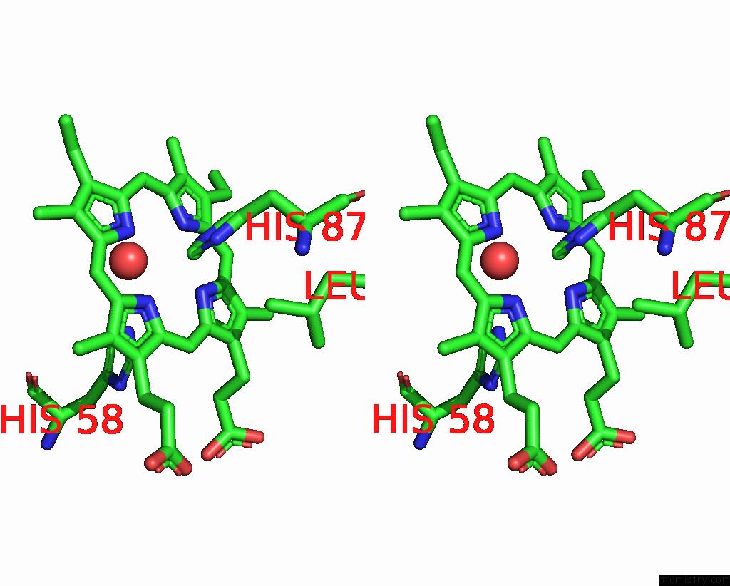
Stereo pair view
Mono view
Stereo pair view
A full contact list of Iron with other atoms in the Fe binding
site number 1 of T-to-Thigh Quaternary Transitions in Human Hemoglobin: ALPHAY140A Deoxy Low-Salt within 5.0Å range:
|
Iron binding site 2 out of 4 in 1y0a
Go back to
Iron binding site 2 out
of 4 in the T-to-Thigh Quaternary Transitions in Human Hemoglobin: ALPHAY140A Deoxy Low-Salt
Mono view
Stereo pair view
Mono view
Stereo pair view
A full contact list of Iron with other atoms in the Fe binding
site number 2 of T-to-Thigh Quaternary Transitions in Human Hemoglobin: ALPHAY140A Deoxy Low-Salt within 5.0Å range:
|
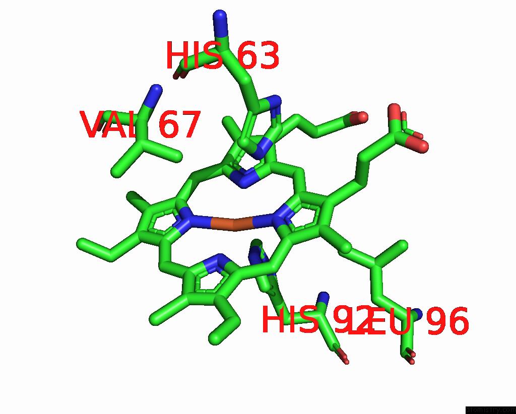
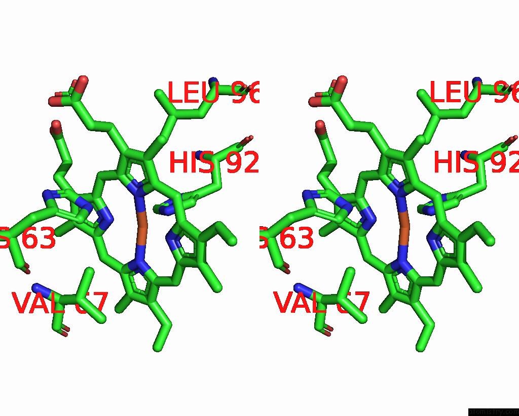
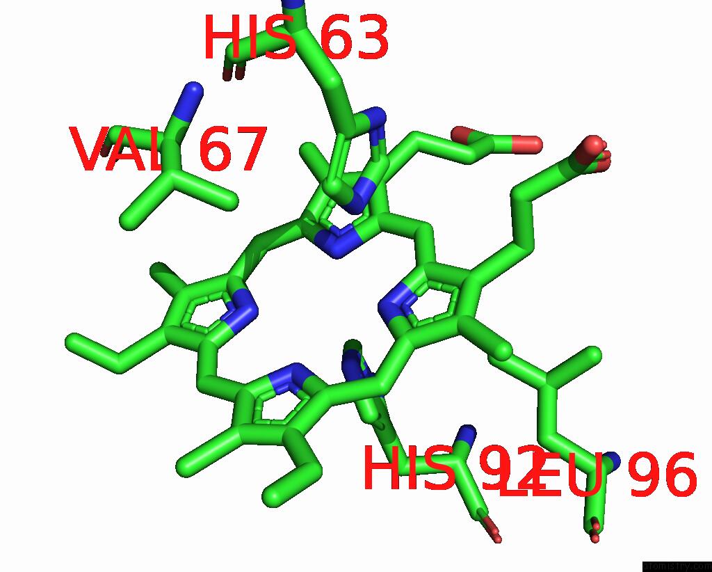
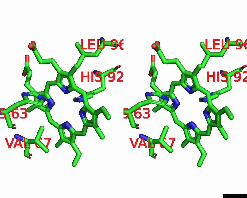
Iron binding site 3 out of 4 in 1y0a
Go back to
Iron binding site 3 out
of 4 in the T-to-Thigh Quaternary Transitions in Human Hemoglobin: ALPHAY140A Deoxy Low-Salt
Mono view
Stereo pair view
Mono view
Stereo pair view
A full contact list of Iron with other atoms in the Fe binding
site number 3 of T-to-Thigh Quaternary Transitions in Human Hemoglobin: ALPHAY140A Deoxy Low-Salt within 5.0Å range:
|
Iron binding site 4 out of 4 in 1y0a
Go back to
Iron binding site 4 out
of 4 in the T-to-Thigh Quaternary Transitions in Human Hemoglobin: ALPHAY140A Deoxy Low-Salt
Mono view
Stereo pair view
Mono view
Stereo pair view
A full contact list of Iron with other atoms in the Fe binding
site number 4 of T-to-Thigh Quaternary Transitions in Human Hemoglobin: ALPHAY140A Deoxy Low-Salt within 5.0Å range:
|
Reference:
J.S.Kavanaugh,
P.H.Rogers,
A.Arnone.
Crystallographic Evidence For A New Ensemble of Ligand-Induced Allosteric Transitions in Hemoglobin: the T-to-T(High) Quaternary Transitions. Biochemistry V. 44 6101 2005.
ISSN: ISSN 0006-2960
PubMed: 15835899
DOI: 10.1021/BI047813A
Page generated: Sat Aug 3 17:09:04 2024
ISSN: ISSN 0006-2960
PubMed: 15835899
DOI: 10.1021/BI047813A
Last articles
Zn in 9J0NZn in 9J0O
Zn in 9J0P
Zn in 9FJX
Zn in 9EKB
Zn in 9C0F
Zn in 9CAH
Zn in 9CH0
Zn in 9CH3
Zn in 9CH1