Iron »
PDB 1yqp-1zj9 »
1yvq »
Iron in PDB 1yvq: The Low Salt (Peg) Crystal Structure of Co Hemoglobin E (BETAE26K) Approaching Physiological pH (pH 7.5)
Protein crystallography data
The structure of The Low Salt (Peg) Crystal Structure of Co Hemoglobin E (BETAE26K) Approaching Physiological pH (pH 7.5), PDB code: 1yvq
was solved by
V.N.Malashkevich,
T.C.Balazs,
S.C.Almo,
R.E.Hirsch,
with X-Ray Crystallography technique. A brief refinement statistics is given in the table below:
| Resolution Low / High (Å) | 23.38 / 1.80 |
| Space group | P 21 21 21 |
| Cell size a, b, c (Å), α, β, γ (°) | 60.621, 96.018, 101.368, 90.00, 90.00, 90.00 |
| R / Rfree (%) | 18.7 / 23 |
Iron Binding Sites:
The binding sites of Iron atom in the The Low Salt (Peg) Crystal Structure of Co Hemoglobin E (BETAE26K) Approaching Physiological pH (pH 7.5)
(pdb code 1yvq). This binding sites where shown within
5.0 Angstroms radius around Iron atom.
In total 4 binding sites of Iron where determined in the The Low Salt (Peg) Crystal Structure of Co Hemoglobin E (BETAE26K) Approaching Physiological pH (pH 7.5), PDB code: 1yvq:
Jump to Iron binding site number: 1; 2; 3; 4;
In total 4 binding sites of Iron where determined in the The Low Salt (Peg) Crystal Structure of Co Hemoglobin E (BETAE26K) Approaching Physiological pH (pH 7.5), PDB code: 1yvq:
Jump to Iron binding site number: 1; 2; 3; 4;
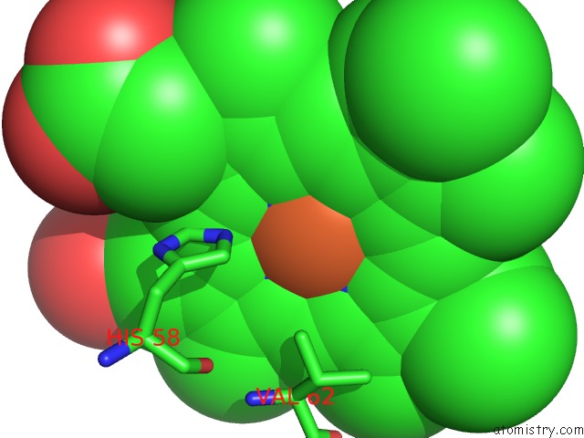
Iron binding site 1 out of 4 in 1yvq
Go back to
Iron binding site 1 out
of 4 in the The Low Salt (Peg) Crystal Structure of Co Hemoglobin E (BETAE26K) Approaching Physiological pH (pH 7.5)
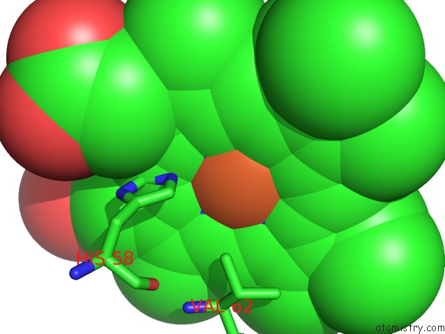
Mono view
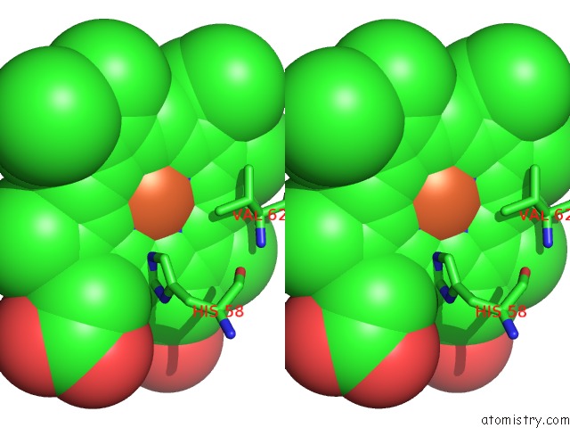
Stereo pair view
Mono view
Stereo pair view
A full contact list of Iron with other atoms in the Fe binding
site number 1 of The Low Salt (Peg) Crystal Structure of Co Hemoglobin E (BETAE26K) Approaching Physiological pH (pH 7.5) within 5.0Å range:
|
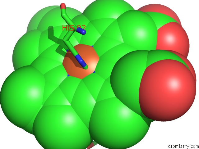
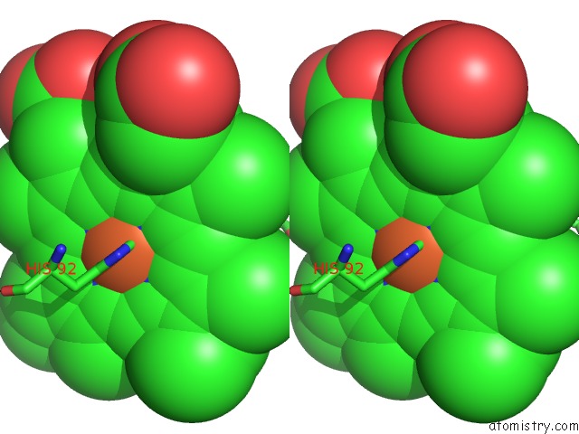
Iron binding site 2 out of 4 in 1yvq
Go back to
Iron binding site 2 out
of 4 in the The Low Salt (Peg) Crystal Structure of Co Hemoglobin E (BETAE26K) Approaching Physiological pH (pH 7.5)
Mono view
Stereo pair view
Mono view
Stereo pair view
A full contact list of Iron with other atoms in the Fe binding
site number 2 of The Low Salt (Peg) Crystal Structure of Co Hemoglobin E (BETAE26K) Approaching Physiological pH (pH 7.5) within 5.0Å range:
|
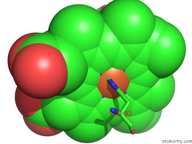
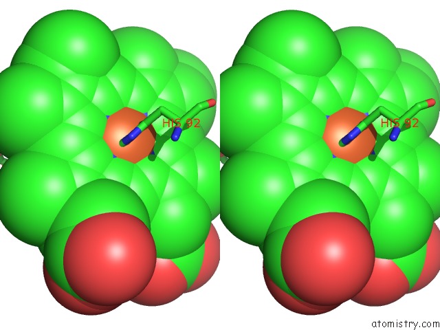
Iron binding site 3 out of 4 in 1yvq
Go back to
Iron binding site 3 out
of 4 in the The Low Salt (Peg) Crystal Structure of Co Hemoglobin E (BETAE26K) Approaching Physiological pH (pH 7.5)
Mono view

Stereo pair view
Mono view
Stereo pair view
A full contact list of Iron with other atoms in the Fe binding
site number 3 of The Low Salt (Peg) Crystal Structure of Co Hemoglobin E (BETAE26K) Approaching Physiological pH (pH 7.5) within 5.0Å range:
|
Iron binding site 4 out of 4 in 1yvq
Go back to
Iron binding site 4 out
of 4 in the The Low Salt (Peg) Crystal Structure of Co Hemoglobin E (BETAE26K) Approaching Physiological pH (pH 7.5)
Mono view
Stereo pair view
Mono view
Stereo pair view
A full contact list of Iron with other atoms in the Fe binding
site number 4 of The Low Salt (Peg) Crystal Structure of Co Hemoglobin E (BETAE26K) Approaching Physiological pH (pH 7.5) within 5.0Å range:
|
Reference:
V.N.Malashkevich,
T.C.Balazs,
S.C.Almo,
R.E.Hirsch.
Crystal Structures of Co Hemoglobin E (BETAE26K) Approaching Physiological pH (pH 7.5) To Be Published.
Page generated: Sat Aug 3 18:04:58 2024
Last articles
Cl in 2XK3Cl in 2XJZ
Cl in 2XJW
Cl in 2XJV
Cl in 2XJU
Cl in 2XJT
Cl in 2XJO
Cl in 2XJS
Cl in 2XJR
Cl in 2XJQ