Iron »
PDB 1yqp-1zj9 »
1z10 »
Iron in PDB 1z10: Crystal Structure of Human Microsomal P450 2A6 with Coumarin Bound
Enzymatic activity of Crystal Structure of Human Microsomal P450 2A6 with Coumarin Bound
All present enzymatic activity of Crystal Structure of Human Microsomal P450 2A6 with Coumarin Bound:
1.14.14.1;
1.14.14.1;
Protein crystallography data
The structure of Crystal Structure of Human Microsomal P450 2A6 with Coumarin Bound, PDB code: 1z10
was solved by
J.K.Yano,
M.H.Hsu,
K.J.Griffin,
C.D.Stout,
E.F.Johnson,
with X-Ray Crystallography technique. A brief refinement statistics is given in the table below:
| Resolution Low / High (Å) | 50.00 / 1.90 |
| Space group | P 1 21 1 |
| Cell size a, b, c (Å), α, β, γ (°) | 70.615, 157.591, 103.541, 90.00, 92.25, 90.00 |
| R / Rfree (%) | 19.4 / 23 |
Iron Binding Sites:
The binding sites of Iron atom in the Crystal Structure of Human Microsomal P450 2A6 with Coumarin Bound
(pdb code 1z10). This binding sites where shown within
5.0 Angstroms radius around Iron atom.
In total 4 binding sites of Iron where determined in the Crystal Structure of Human Microsomal P450 2A6 with Coumarin Bound, PDB code: 1z10:
Jump to Iron binding site number: 1; 2; 3; 4;
In total 4 binding sites of Iron where determined in the Crystal Structure of Human Microsomal P450 2A6 with Coumarin Bound, PDB code: 1z10:
Jump to Iron binding site number: 1; 2; 3; 4;
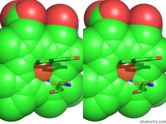
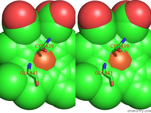
Iron binding site 1 out of 4 in 1z10
Go back to
Iron binding site 1 out
of 4 in the Crystal Structure of Human Microsomal P450 2A6 with Coumarin Bound

Mono view
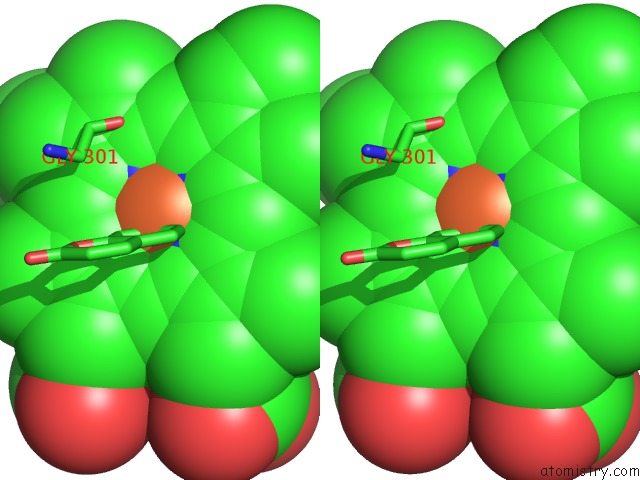
Stereo pair view
Mono view
Stereo pair view
A full contact list of Iron with other atoms in the Fe binding
site number 1 of Crystal Structure of Human Microsomal P450 2A6 with Coumarin Bound within 5.0Å range:
|
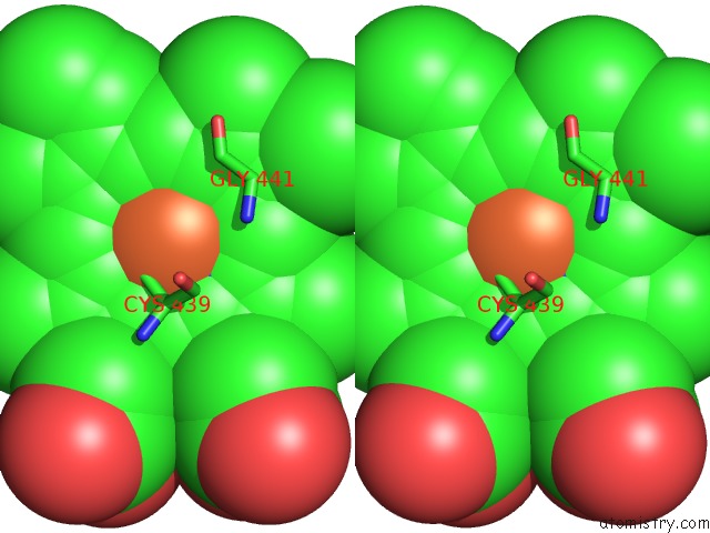
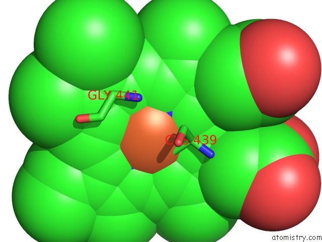
Iron binding site 2 out of 4 in 1z10
Go back to
Iron binding site 2 out
of 4 in the Crystal Structure of Human Microsomal P450 2A6 with Coumarin Bound

Mono view
Stereo pair view
Mono view
Stereo pair view
A full contact list of Iron with other atoms in the Fe binding
site number 2 of Crystal Structure of Human Microsomal P450 2A6 with Coumarin Bound within 5.0Å range:
|
Iron binding site 3 out of 4 in 1z10
Go back to
Iron binding site 3 out
of 4 in the Crystal Structure of Human Microsomal P450 2A6 with Coumarin Bound

Mono view
Stereo pair view
Mono view
Stereo pair view
A full contact list of Iron with other atoms in the Fe binding
site number 3 of Crystal Structure of Human Microsomal P450 2A6 with Coumarin Bound within 5.0Å range:
|
Iron binding site 4 out of 4 in 1z10
Go back to
Iron binding site 4 out
of 4 in the Crystal Structure of Human Microsomal P450 2A6 with Coumarin Bound
Mono view
Stereo pair view
Mono view
Stereo pair view
A full contact list of Iron with other atoms in the Fe binding
site number 4 of Crystal Structure of Human Microsomal P450 2A6 with Coumarin Bound within 5.0Å range:
|
Reference:
J.K.Yano,
M.H.Hsu,
K.J.Griffin,
C.D.Stout,
E.F.Johnson.
Structures of Human Microsomal Cytochrome P450 2A6 Complexed with Coumarin and Methoxsalen Nat.Struct.Mol.Biol. V. 12 822 2005.
ISSN: ISSN 1545-9993
PubMed: 16086027
DOI: 10.1038/NSMB971
Page generated: Wed Jul 16 23:05:06 2025
ISSN: ISSN 1545-9993
PubMed: 16086027
DOI: 10.1038/NSMB971
Last articles
Fe in 2YXOFe in 2YRS
Fe in 2YXC
Fe in 2YNM
Fe in 2YVJ
Fe in 2YP1
Fe in 2YU2
Fe in 2YU1
Fe in 2YQB
Fe in 2YOO