Iron »
PDB 1yqp-1zj9 »
1zby »
Iron in PDB 1zby: High-Resolution Crystal Structure of Native (Resting) Cytochrome C Peroxidase (Ccp)
Enzymatic activity of High-Resolution Crystal Structure of Native (Resting) Cytochrome C Peroxidase (Ccp)
All present enzymatic activity of High-Resolution Crystal Structure of Native (Resting) Cytochrome C Peroxidase (Ccp):
1.11.1.5;
1.11.1.5;
Protein crystallography data
The structure of High-Resolution Crystal Structure of Native (Resting) Cytochrome C Peroxidase (Ccp), PDB code: 1zby
was solved by
C.A.Bonagura,
B.Bhaskar,
H.Shimizu,
H.Li,
M.Sundaramoorthy,
D.E.Mcree,
D.B.Goodin,
T.L.Poulos,
with X-Ray Crystallography technique. A brief refinement statistics is given in the table below:
| Resolution Low / High (Å) | 100.00 / 1.20 |
| Space group | P 21 21 21 |
| Cell size a, b, c (Å), α, β, γ (°) | 106.673, 75.513, 50.994, 90.00, 90.00, 90.00 |
| R / Rfree (%) | 11.3 / 14.9 |
Iron Binding Sites:
The binding sites of Iron atom in the High-Resolution Crystal Structure of Native (Resting) Cytochrome C Peroxidase (Ccp)
(pdb code 1zby). This binding sites where shown within
5.0 Angstroms radius around Iron atom.
In total only one binding site of Iron was determined in the High-Resolution Crystal Structure of Native (Resting) Cytochrome C Peroxidase (Ccp), PDB code: 1zby:
In total only one binding site of Iron was determined in the High-Resolution Crystal Structure of Native (Resting) Cytochrome C Peroxidase (Ccp), PDB code: 1zby:
Iron binding site 1 out of 1 in 1zby
Go back to
Iron binding site 1 out
of 1 in the High-Resolution Crystal Structure of Native (Resting) Cytochrome C Peroxidase (Ccp)
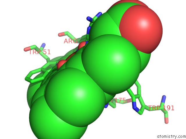
Mono view
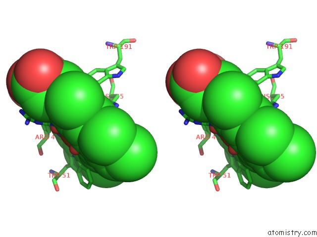
Stereo pair view
Mono view
Stereo pair view
A full contact list of Iron with other atoms in the Fe binding
site number 1 of High-Resolution Crystal Structure of Native (Resting) Cytochrome C Peroxidase (Ccp) within 5.0Å range:
|
Reference:
C.A.Bonagura,
B.Bhaskar,
H.Shimizu,
H.Li,
M.Sundaramoorthy,
D.E.Mcree,
D.B.Goodin,
T.L.Poulos.
High-Resolution Crystal Structures and Spectroscopy of Native and Compound I Cytochrome C Peroxidase Biochemistry V. 42 5600 2003.
ISSN: ISSN 0006-2960
PubMed: 12741816
DOI: 10.1021/BI034058C
Page generated: Sat Aug 3 18:16:12 2024
ISSN: ISSN 0006-2960
PubMed: 12741816
DOI: 10.1021/BI034058C
Last articles
Zn in 9MJ5Zn in 9HNW
Zn in 9G0L
Zn in 9FNE
Zn in 9DZN
Zn in 9E0I
Zn in 9D32
Zn in 9DAK
Zn in 8ZXC
Zn in 8ZUF