Iron »
PDB 1yqp-1zj9 »
1zgn »
Iron in PDB 1zgn: Crystal Structure of the Glutathione Transferase Pi in Complex with Dinitrosyl-Diglutathionyl Iron Complex
Enzymatic activity of Crystal Structure of the Glutathione Transferase Pi in Complex with Dinitrosyl-Diglutathionyl Iron Complex
All present enzymatic activity of Crystal Structure of the Glutathione Transferase Pi in Complex with Dinitrosyl-Diglutathionyl Iron Complex:
2.5.1.18;
2.5.1.18;
Protein crystallography data
The structure of Crystal Structure of the Glutathione Transferase Pi in Complex with Dinitrosyl-Diglutathionyl Iron Complex, PDB code: 1zgn
was solved by
L.J.Parker,
J.J.Adams,
M.W.Parker,
with X-Ray Crystallography technique. A brief refinement statistics is given in the table below:
| Resolution Low / High (Å) | 50.00 / 2.10 |
| Space group | C 1 2 1 |
| Cell size a, b, c (Å), α, β, γ (°) | 76.219, 89.759, 68.713, 90.00, 97.63, 90.00 |
| R / Rfree (%) | 18.2 / 24.4 |
Iron Binding Sites:
The binding sites of Iron atom in the Crystal Structure of the Glutathione Transferase Pi in Complex with Dinitrosyl-Diglutathionyl Iron Complex
(pdb code 1zgn). This binding sites where shown within
5.0 Angstroms radius around Iron atom.
In total 2 binding sites of Iron where determined in the Crystal Structure of the Glutathione Transferase Pi in Complex with Dinitrosyl-Diglutathionyl Iron Complex, PDB code: 1zgn:
Jump to Iron binding site number: 1; 2;
In total 2 binding sites of Iron where determined in the Crystal Structure of the Glutathione Transferase Pi in Complex with Dinitrosyl-Diglutathionyl Iron Complex, PDB code: 1zgn:
Jump to Iron binding site number: 1; 2;
Iron binding site 1 out of 2 in 1zgn
Go back to
Iron binding site 1 out
of 2 in the Crystal Structure of the Glutathione Transferase Pi in Complex with Dinitrosyl-Diglutathionyl Iron Complex
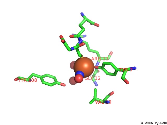
Mono view
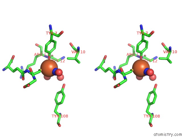
Stereo pair view
Mono view
Stereo pair view
A full contact list of Iron with other atoms in the Fe binding
site number 1 of Crystal Structure of the Glutathione Transferase Pi in Complex with Dinitrosyl-Diglutathionyl Iron Complex within 5.0Å range:
|
Iron binding site 2 out of 2 in 1zgn
Go back to
Iron binding site 2 out
of 2 in the Crystal Structure of the Glutathione Transferase Pi in Complex with Dinitrosyl-Diglutathionyl Iron Complex
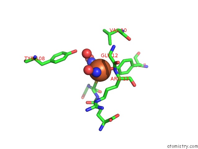
Mono view
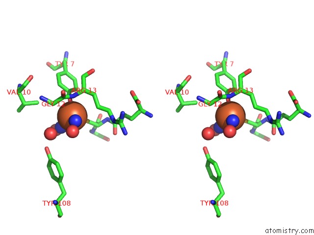
Stereo pair view
Mono view
Stereo pair view
A full contact list of Iron with other atoms in the Fe binding
site number 2 of Crystal Structure of the Glutathione Transferase Pi in Complex with Dinitrosyl-Diglutathionyl Iron Complex within 5.0Å range:
|
Reference:
E.Cesareo,
L.J.Parker,
J.Z.Pedersen,
M.Nuccetelli,
A.P.Mazzetti,
A.Pastore,
G.Federici,
A.M.Caccuri,
G.Ricci,
J.J.Adams,
M.W.Parker,
M.L.Bello.
Nitrosylation of Human Glutathione Transferase P1-1 with Dinitrosyl Diglutathionyl Iron Complex in Vitro and in Vivo J.Biol.Chem. V. 280 42172 2005.
ISSN: ISSN 0021-9258
PubMed: 16195232
DOI: 10.1074/JBC.M507916200
Page generated: Sat Aug 3 18:17:23 2024
ISSN: ISSN 0021-9258
PubMed: 16195232
DOI: 10.1074/JBC.M507916200
Last articles
Zn in 9MJ5Zn in 9HNW
Zn in 9G0L
Zn in 9FNE
Zn in 9DZN
Zn in 9E0I
Zn in 9D32
Zn in 9DAK
Zn in 8ZXC
Zn in 8ZUF